Chromatin and Chromatid
Chromatin and Chromatid are key components in organizing, packaging, and transmitting genetic material within cells. While they share similarities in structure, each offers distinct properties which play unique roles in cellular processes.
Chromatin is an intricate structure formed of DNA and proteins found within eukaryotic cell nuclei. The main role of chromatin formation is packaging DNA for efficient storage within limited nuclear space; histone proteins play an essential part here, protecting DNA from damage while simultaneously controlling gene expression and providing access to genetic information.
Chromatin does not remain static throughout its transformations within cell cycles, yet continuously undergoes dynamic shifts that enable gene transcription and protein synthesis to take place. When not actively dividing, interphase-era chromatin transitions to more relaxed states known as euchromatin that allow access to genetic material necessary for gene transcription or protein synthesis.
During cell division, this same state becomes compacted again to form visible chromosomes that can be seen under the microscope. Chromatid refers to one of two identical copies of a replicated chromosome and is formed during the S phase of cell division when DNA replication takes place.
Each sister chromatid joins together at their centromere region before being passed along during cell division to daughter cells as genetic material is accurately distributed across them by spindle fibers during mitosis resulting in two individual copies being attached at opposite poles of each daughter cell, eventually giving rise to two individual chromosomes within each daughter cell.
Importance of understanding the difference between chromatin and chromatid
Understanding the difference between chromatin and chromatid is vital to understanding cell biology and genetics, for several key reasons:
- Cellular processes: Chromatin and chromatid both play key roles in various cellular processes, from gene expression regulation, DNA packaging, and organization throughout cell division to accurate chromosome segregation during division. Understanding their respective roles is integral for understanding processes like transcription, DNA replication, and mitosis.
- Genetic Stability: Chromatin and chromatid are fundamental to maintaining genetic stability. Their structure and modifications can dramatically impact gene access and regulation, potentially leading to changes in cell function or even disease development. Proper segregation of chromatids during cell division is vital in order to avoid errors such as chromosomal aberrations and aneuploidy which could have severe implications such as genetic disorders or cancer. understanding how each of them works helps shed light on mechanisms involved with maintaining genetic stability.
- Research and Therapeutics: Studies in molecular biology and genetics depend heavily on differentiating between chromatin and chromatid structures for accurate investigation of their interactions. ChIP and 3C techniques are employed to probe these structures further while targeting modifications or proteins associated with them can offer therapeutic interventions in various diseases, including cancer; distinguishing chromatin from chromatid is crucial in creating effective treatment plans.
- Education and scientific communication: Understanding scientific concepts clearly is of utmost importance when it comes to education and scientific research. For example, understanding the differences between chromatin and chromatid helps avoid confusion and ensures precise communication among researchers, educators, and students and knowledge transfer between researchers. Furthermore, accurate interpretation of literature facilitates effective knowledge transfer.
Structure of Chromatin
The chromatin structure is intricate and comprises DNA molecules that are tightly wrapped around proteins referred to as histones. The chromatin’s basic component is the nucleosome. It is made up of DNA wrapped in a central core of histone proteins.
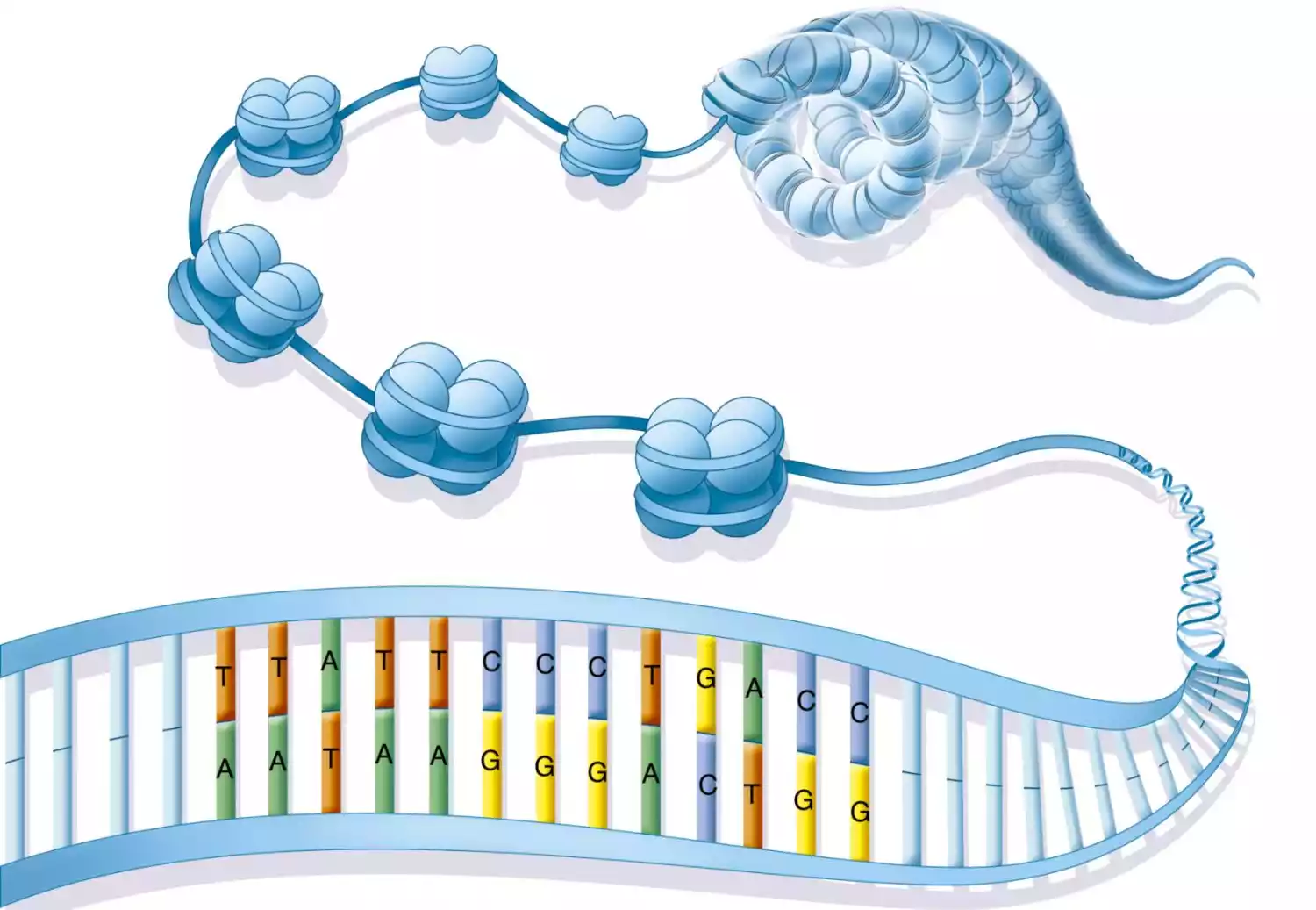
The histones are tiny positively charged protein molecules that are able to interact with negatively charged DNA helping stabilize and regulate the structure of chromatin. Each nucleosome contains around 150 base pairs of DNA wrapped around an octamer made of histone proteins. It comprises duplicates of all four core histones: H2A and He2 and H3. The histone octamer acts as a structural scaffold and DNA is wrapped around it during the left-hand superhelical turn. The wrapping leads to the creation of a compact and well-organized structure.
Multiple nucleosomes are linked with DNA by a short stretch known as linker DNA. these nucleosomes together with linker DNA create an “beads-on-a-string” structure. This structure may be further folded and coil, leading to greater levels of compaction of chromatin.
There are two major types of chromatin compaction, known as heterochromatin and Euchromatin. Euchromatin is less condensed and is connected to genes that are active, giving access to DNA that is used to create transcriptional machinery. Heterochromatin On the contrary is more compact and is typically is associated with silenced or inactive genes.
Alongside histones, the structure of chromatin is also affected by non-histone proteins like chromatin remodelers, structural proteins, and modifiers. The structure of Chromatin is constantly changing and is able to change to accommodate a variety of cell functions like the expression of genes, replication of DNA and repair of DNA. The precise arrangement and the compaction of chromatin is essential to good gene regulation and the preservation of genome integrity.
What common types of chromatin are:
There are 2 major kinds of chromatin: heterochromatin and euchromatin.
- Euchromatin: Euchromatin refers to the less condensed, but more active transcriptionally active version of the chromatin. It’s distinguished by the more open structure which permits access to the DNA and enhances the expression of genes. Euchromatin is often connected to genes that are being actively transcribed and genomic regions that are involved in regulatory processes. It is seen as an expansive and less stained region when examined under a microscope.
- Heterochromatin: Heterochromatin is the extremely condensed and inactive transcription version of the chromatin. It is densely packed and appears to be a more densely stained and condensed region under a microscope. Heterochromatin tends to be abundant in repetitive DNA sequences as well as regions of the genome that do not undergo active transcription.
It is an important factor in ensuring the stability of the genome by shielding the genome from harming elements, and also regulating gene expression by blocking genes in certain cell or developmental contexts.
There are subtypes and variations of chromatin that may exist. For instance, facultative heterochromatin refers to regions of the genome that are able to switch between euchromatin or heterochromatin state, based on the stage of development or the cellular context. Constitutive heterochromatin is a term used to describe regions that are in a condensed condition throughout the life cycle of cells.
Structure of Chromatid
A chromatid is a condensed and tightly wound structure that represents one of the two identical copies of a replicated chromosome, and forms during the S phase of the cell cycle during DNA replication.
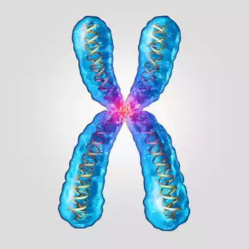
Each chromatid contains an exact replica of its respective original chromosome’s DNA molecule, held together at its centromere by bonds known as sister chromatid pairs and also serving as attachment sites for spindle fibers during cell division.
Chromatids are similar to chromosomes in structure and function; the main distinction lies in that chromatids are temporary structures produced during cell cycle progression that become highly condensed and visible under a microscope. Chromatid condensation occurs through the coiling and folding of replicated DNA molecules which is assisted by proteins to maintain the structure and stability of each chromatid.
Chromatids play an integral part in cell division by ensuring the accurate distribution of genetic material to daughter cells. During mitosis, sister chromatids of each chromosome separate and are pulled apart using spindle fibers; once separated they each become an individual chromosome in each daughter cell.
Once cell division has concluded, chromatids uncoil and become less condensed, returning to a more extended and relaxed chromatin state for use during the interphase of the cell cycle.
Chromatids are condensed and tightly wound structures that represent one of two identical copies of a replicated chromosome. Chromatids form during DNA replication and play an essential role in ensuring the accurate distribution of genetic material during cell division.
What common types of Chromatid are:
There aren’t specific “types” of chromatids since chromatids are the duplicated copies of a chromosome throughout a cell’s cycle. There are a variety of phases and conditions that can be that are associated with chromatids.
- Replicated Chromatids: When DNA replication takes place during the S phase of the cell cycle it leads to the formation of two copies identical to the DNA chromosome. The duplicated copies are called sister chromatids. They are bonded by the centromere region until they split when cells divide.
- Condensed Chromatids: Chromomatids undergo condensation and coiling, making them extremely compact and easily visible under microscopes when cells divide. Condensed states allow the proper segregation and separation of genetic material in mitosis and meiosis.
It is crucial to remember that the phrase “chromatid” is commonly used to refer to the repeated copies of a single chromosome in cell division, particularly during meiosis or mitosis. After chromatids are separated and dispersed among daughter cells transform into individual chromosomes.
Difference Between Chromatin and Chromatid
The key differences between chromatin and chromatid can be summarized as follows:
Definition:
- Chromatin : Chromatin is the DNA complex and the associated proteins that are located in the nucleus of cells.
- Chromatid: A chromatid can be described as one of 2 identical copies of the duplicated chromosome.
Structure:
- Chromatin: Chromatin also known as Chromatin, is a fairly large as well as loosely packed DNA protein complex. It is composed of DNA encased in histone proteins. They form nucleosomes as well as chromatin fibers.
- Chromatids: A chromatid is an encapsulated and tightly coiled structure created in the course of DNA replication. It represents one of 2 identical copies of the duplicated chromosome. It is visible under microscopes during the division of cells.
Formation:
- Chromatin: Chromatin exists throughout the cell cycle, and serves in the state of default for DNA, even when it’s not actively replicating or dividing.
- Chromatids: Chromatids develop in the S phase of the cell cycle, when DNA replication occurs. Each replicated chromosome leads two sister chromatids, held together by the centromere.
Role:
- Chromatin: Chromatin is an essential role in the process of storing and organizing DNA inside the nucleus. It aids in controlling gene expression by regulating access to genetic information.
- Chromatids: Chromatids play a key role associated with cell division. When meiosis or mitosis occurs the chromatids split and move towards opposite poles of the cell, ensuring precise dispersal of DNA to the daughter cells.
The similarity between Chromatin and Chromatid
- DNA Composition: Both chromatins, as well as chromatids, have DNA as their main component. Chromatin and Chromatid DNA molecules are made up of Nucleotides that are arranged in a certain sequence that encodes DNA’s genetic data in the organism.
- Replication: Chromatin, Chromatid, and Chromatid play a role in the process of DNA replication. In the S phase of the cell cycle DNA replication occurs, which results in the creation of duplicate copies of chromosomes. These duplicated copies are known as sister chromatids that are bonded by the centromere.
- Histone Proteins: Both the chromatin as well as the chromatid are related to histone proteins. Histones play an important role in the organization and packaging of DNA. In both instances, DNA is encased in histone proteins and forms nucleosomes. They are the chromatin’s basic structure.
- Packaging: Chromatin and the chromatid undergo structural modifications to allow the efficient packaging of DNA. While chromatin tends to be more flexible and easy to access, both chromatin and the chromatid are prone to condensation and compaction to ensure proper chromosome organization and segregation in cell division.
- The accessibility of DNA: Chromatin as well as the chromatid undergo changes in their structure in order to regulate accessibility to DNA. Chromatin is able to transition into open (euchromatin) as well as less compact (heterochromatin) forms, whereas the chromatid’s condensation in cell division is essential to ensure the segregation of chromosomes.
Summary
Chromatin and Chromatid are essential components of the intricate DNA packaging mechanisms within our cells. The dynamic relationship between chromatin and chromatid plays a fundamental role in cellular processes, including cell division and gene regulation. Understanding these molecular intricacies is crucial for unlocking the mysteries of genetics and its impact on human health and disease.