When studying parasitic organisms, it becomes crucial to understand their different life stages. Cyst and Trophozoite are two such stages that exhibit unique properties and behaviors. Let’s explore each of them in detail.
Definition of Cyst
The term “cyst” refers to a sac-like structure or a swollen pocket of tissue that may be found either on or within the body inside the human body. It’s typically comprised of semi-solid or fluid substances or gas. Cysts may differ in size, ranging from microscopic to massive forms which can be felt or observed. They can form in different organs, tissues, and body cavities, such as but not only Ovaries, the skin liver, kidneys, as well as bones.
Cysts may develop for various causes, including blocked ducts, infections or developmental anomalies. They may appear benign (non-cancerous) or malignant (cancerous). The benign cysts are generally harmless and don’t necessitate treatment until they trigger a symptom or cause complications. Malignant cysts, on the contrary, can be cancerous and require medical treatment.
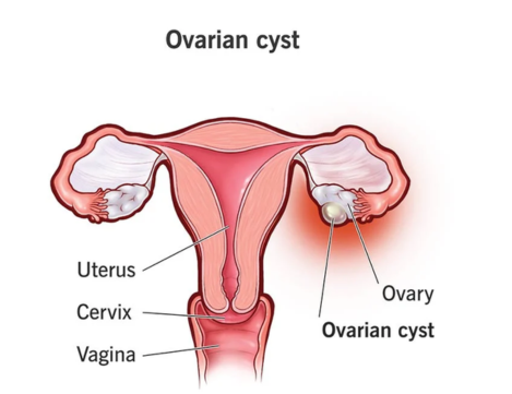
The appearance of cysts may differ depending on their origin and location. The most common kinds of cysts are sebaceous cysts (filled with oily substance) and the ovarian cysts (develop within the Ovaries) and Ganglion cysts (fluid-filled sacs located near joints or tendon). The diagnosis of a cyst typically requires physical examination and testing for imaging (such such as ultrasound and MRI) as well as, occasionally an examination of the tissue or fluid (biopsy) to further analyze.
The treatment of cysts is based on the factors involved, Including the size, location, and symptoms and whether they’re malignant or Benign. Treatment options could include medication, observation, surgical removal, drainage or other procedures that are specialized. It is essential to speak with an expert in healthcare for the appropriate evaluation and treatment of cysts.
Characteristics of Cysts
Cysts have distinct characteristics that differentiate these from the other structures or anomalies within the body.
Here are a few of the key characteristic characteristics that are associated with cysts:
- A sac-like structure: Cysts are generally enclosed in the sac-like structure composed of epithelial cells and connective tissue. This sac serves as a barrier that protects the cyst. It also is the home for what is inside the cyst.
- Semi-solid, fluid or gas-filled: Cysts can contain different substances, such as fluids semi-solid material, fluid, or gas. Contents of cysts vary based on its nature and location.
- Cysts of different sizes: They can vary in size, from tiny to massive formations that may be observable or felt. The size of the cyst can affect symptoms, complications and treatments options.
- Smooth or irregular surface: outside of a cyst could appear smooth or uneven depending on the area it is located along with the extent of swelling or inflammation.
- Encapsulation: Cysts tend to be sealed, which means that they are enclosed by a separate barrier or wall. The cyst’s membrane is able to separate it from surrounding tissues, and also helps keep its contents contained.
- Slow growth: Cysts typically expand gradually in time. The rate at which they grow can depend on aspects like the kind of cyst, the underlying reason, and other specific factors.
- Malignant or benign: Cysts can be benign (non-cancerous) or malignant (cancerous). The benign cysts are generally harmless and don’t expand to other parts in the body. The malignant cysts, on other hand, possess the potential of invading surrounding tissues and cause metastasis.
- Acquired or developed: Cysts are either developing, meaning that they are formed during the development of embryos or in young life, or acquired, forming later in life as a result of circumstances like blockages, infections, or other pathological conditions.
- Symptoms and complications: The symptoms and complications vary based on the size or location as well as their contents, cysts can or might not cause symptoms. Some cysts are not symptomatic, others could cause swelling, pain, infection or other problems in the event that they expand, rupture or develop infection.
- Methods to diagnose: Cysts can be typically diagnosed by physical examination and scans (such such as ultrasound MRI as well as CT scans) as well as, occasionally samples of fluid or tissue (biopsy) to further analyze.
Bear in mind that, while these characteristics often associated with cysts can vary depending on their type and location. A consultation with a doctor is essential for accurate diagnosis and proper treatment of cysts.
Structure and Form
The shape and structure of cysts vary based on the nature as well as the location. But, there are a few common features which can be described
- Outer membrane or wall: Cysts are usually enclosed in an outer membrane, or wall, that isolates them from other tissues. Membranes consist of epithelial cells and connective tissues; thickness can differ based on composition and size.
- The inner lining: Within of the cyst could exist an internal lining made up of cells that are specialized. The lining could release fluids as well as other components that aid in the content inside the cyst.
- Contents: Cysts can contain liquid, semi-solid or gas. The exact contents may differ dependent on the kind of cyst. For instance, some cysts might contain serous or clear fluid, while other cysts may contain pus-like, thicker material or even solid parts.
- Shape and size: Cysts vary in size, from very tiny microscopically-sized structures to larger, more complex ones which are visible or palpable. The shape of a cyst could differ and include oval or round, while others with irregular or lobulated forms.
- Consistency: A cyst’s consistency cyst is based on its texture or the feel. Some cysts feel soft or sluggish and have an ethereal or fluid-like sensation when they are touched. Other cysts may feel hard or solid depending on the type of the contents and the growth of any tissue.
- Surrounding tissues: Cysts are known to cause different reactions from the surrounding tissues. In certain instances an occurrence of cysts can result in displacement or compression of nearby structures. In addition the surrounding tissues could show indications of inflammation or an immune response when the cyst is damaged or ruptures.
That cyst formation and structure depend on many variables; such as their cause, individual variation and organ/tissue-specific features. A thorough imaging technique or a microscopic exam might be required to identify the structure and shape of a particular cyst.
Function and Survival
Cysts perform a variety of functions and have mechanisms for survival that allow them to stay within the body and in external environments.
Here are a few aspects that relate to the purpose and existence of cysts:
- Encapsulation and protection: The outer membrane, (or wall) of the cyst gives protection as well as encapsulates its contents, thus separating it from the surrounding tissues or the outside surrounding environment. Encapsulation can prevent destruction or damage to the contents of the cyst.
- Dormant condition: Cysts typically go into a quiescent or dormant state. This permits them to live in difficult conditions, like the lack of nutrients or extreme temperatures. dangerous conditions. When they are in dormancy the energy production in the cyst gets drastically decreased, which helps to conserve the energy as well as resources.
- Resilience to adverse conditions: Cysts are resistant to circumstances that could jeopardize their existence. The membrane that surrounds the outside inside the cyst together with its dormant state could offer protection against chemical, physical or immune-mediated attacks. This helps cysts remain in hostile environments or in the immune system of the host.
- Survival and transmission outside of the host: For certain parasites and parasites cysts are an vital role in transmitting into new hosts. Cysts can leak to the outside world via the secretions of feces or through other methods. Through the formation of cysts, parasites boost the likelihood of surviving outside the host, which allows them to live and eventually infect new hosts via a variety of methods, including the ingestion process or vector transmission.
- Persistence in the body: Certain cysts are able to remain within the body for long time. They could remain dormant, or expand slowly, eventually evading the immune system and possibly cause persistent infections. This persistency contributes to the persistence of the parasite in the host.
- Strategies to survive treatment: Cysts may have survival strategies that render them intolerant to specific treatments. For instance, certain cysts possess mechanisms to avoid or resist antimicrobials which allows them to live and possibly cause recurrences as well as treatment failing.
- Facilitating the development of parasites and their development of the parasite’s lifecycle: Cysts are essential parts of the life cycle of parasites. They can serve as a stage to allow reproduction, development or transition to the other forms of the parasite like trophozoites or larvae. In facilitating the life cycle, cysts aid to the survival and perpetuation of the parasite’s species.
As it varies depending on their species and host parasite or disease, cyst functions and mechanisms for survival vary considerably. Understanding these mechanisms is vital for a proper diagnosis, treatment and prevention strategies that target cyst-forming parasites as well as cyst-related illnesses.
Definition of Trophozoite
A trophozoite is a mobile and active feeding stage in the development of a few protozoan parasites. It is the active or vegetative type of parasite that can grow in size, replication, and even leading to diseases within the host. Trophozoites are usually distinguished through their capability to move around and feed on host tissue as well as other materials.
“Thrombozite” or “trophozoite” is commonly used to describe protozoan parasites. Particularly, they are of the phylum Apicomplexa or the amoebae group. They have complicated life cycles with different stages and the trophozoite phase is generally closely associated with the invasive pathogenic stage of the infection.
Trophozoites have distinct characteristics based on the particular parasite species and the interaction and host.
The most common characteristics of trophozoites are:
- Motility: Trophozoites typically are capable of active movement through structures like flagella and cilia as well as pseudopodia or mechanisms for gliding. Their motility permits them to move and enter the tissues of their hosts or search for nutrients.
- Food and metabolism: Trophozoites feed on tissues, cells and bodily fluids as well as other organic materials. They are able to produce enzymes and organelles required for the acquisition of nutrients and energy production via processes such as pinocytosis and phagocytosis as well as absorption.
- Growth and replication: The Trophozoites are able to reproduce and multiply inside the body of the host. The cells they infect undergo division as well as an expansion which results in increased parasite load as well as the potential for damage to host tissues.
- Pathogenicity: Trophozoites could cause illness and have pathogenic consequences in the host. They could produce toxins, enzymes or inflammatory reactions which can cause inflammation, tissue damage and the emergence of clinical signs.
- The susceptibility to host immune response: Trophozoites can interact with the immune system of the host and can trigger an attack against invading parasites. Trophozoites can utilize strategies to avoid or modify the immune response of the host and allow them to remain and cause disease.
A trophozoite’s presence within the diagnostic samples, like stool, tissues or bodily fluids can be a sign that there is an active disease caused by the protozoan parasite. Identification and detection of trophozoites is essential for determining the correct diagnosis, ensuring proper treatment as well as knowing the cause of the parasite’s infection.
Characteristics of Trophozoites
- Motility: Trophozoites tend to be mobile and have a variety of mechanisms of motion. This may include the use of flagella pseudopodia, cilia (temporary extension of cell membranes) or gliding motion. The precise method of motion is dependent on the species of parasite.
- Cellular morphology: Trophozoites typically possess distinct cellular morphologies and can differ greatly dependent on the parasite. They can be amoeboid or amorphous in shape, showing an elongated and flexible shape or possess a more distinct and structured cell form.
- The metabolism and feeding: Trophozoites are active in feeding and metabolizing inside the body of their host. They obtain nutrients by taking in organic material or engulfing it including tissues, host cells and bodily fluids. Trophozoites have specialized organelles, metabolic pathways, and enzymes to help them meet their energy needs and development.
- Nucleus and cytoplasmic features: The cytoplasm and nucleus are the most prominent features. Trophozoites generally have a distinct nucleus that houses DNA of parasites. The cytoplasm in trophozoites could be home to various organelles, including mitochondria, Golgi apparatus and endoplasmic-reticulum, which play a vital role in cells’ functions.
- Replication and proliferative capacity: Trophozoites are able to reproduce and multiply in the human host. Cell division is a process they undergo which can be accomplished through binary fission or other methods of asexual reproduction. This can lead to a rise in parasite populations.
- Pathogenicity: Trophozoites are pathogenic and can cause illness within the host. They might have virulence-related factors, like enzymes, toxins, or adhesive structures that can cause inflammation, tissue damage and the emergence of clinical signs.
- The host tissue invaders: Trophozoites possess the ability to infiltrate tissue of the host, penetrating epithelial barriers, and possibly infiltrating the bloodstream and other organs. Their invading nature permits them to spread an infection and aid in the cause and progression of disease.
- Involvement with immune systems: Trophozoites have the capacity to interact with and trigger an immune response against parasites that invade. This response might involve various elements such as phagocytes, cytokines or antibodies; while trophozoites could employ mechanisms that alter or block such responses in order to survive and spread disease in their host host.
It is important to recognize that the trophozoites’ features are largely dependent on the parasite species that is in question. A thorough microscopic examination as well as molecular techniques and other diagnostic techniques are usually needed to determine and identify trophozoites in the clinical or research setting.
Structure and Form
The shape and structure of trophozoites vary based on the protozoan parasite type.
There are some common features and structures that are common to trophozoites:
- Cell membrane: Trophozoites consist of cells with cells covered with cell membranes that protect and surround their cytoplasmic components within. This barrier acts to safeguard their contents as well as regulate how substances move both into and out of its domain.
- Cytoplasm: the cytoplasm within the trophozoites has a myriad of organelles and structures that play a role in cellular processes. They could comprise mitochondria, the nucleus, Golgi apparatus and endoplasmic retina and cytoskeletal components.
- Nucleus: Trophozoites generally have a distinct nucleus which includes DNA of parasites. The nucleus controls cell processes and also includes the DNA needed to replicate and protein synthesizing.
- Organelles: Trophozoites can have various organelles based on the parasite species they are in. For instance, certain species of trophozoites could possess mitochondria that produce energy as well as the Golgi apparatus for processing protein and secretion and an endoplasmic-reticulum to facilitate protein synthesis.
- Pseudopodia and flagella: Certain trophozoites, like ones belonging to amoeboid group or flagellated protozoa, possess specialized structures to move. Amoeboid trophozoites are able to stretch and retract their pseudopodia that are temporary extensions of cell membranes to aid in mobility. The flagellated trophozoites are equipped with flagella with a whip shape which propel them through the surrounding fluid.
- Cytoskeleton: Trophozoites typically have a cytoskeleton which gives structural support and helps keep the shape of the cell. The cytoskeleton comprises filaments of protein, like tubulin and actin, which help in cell stability, movement and change in shape.
- Surface characteristics: Trophozoites could have particular surface characteristics which aid in the attachment or invading host tissue. These may include flagella, cilia, pseudopodia, or other adhesive structures that are located on the cell’s surface.
- Shape and size: Shape and size of trophozoites vary with parasite species; from being small and round, to larger irregularly-shaped formations. Their shapes can change according to environmental conditions or at various development stages in parasite life cycles.
Recognizing that trophozoites come in various structures and forms is crucial in understanding protozoan parasite diversity as well as adaption to diverse host environments. Microscopy, advanced imaging techniques and molecular analysis are typically employed for characterizing structural traits of trophozoites.
Function and Activity
Trophozoites play an essential part in protozoan parasite life cycles and pathology, providing specific functions and actions which allow them to live their lifecycle of reproduction, multiplication and possible infection of hosts.
Here are a few key roles and functions played by trophozoites:
- Nutrition and feeding: Trophozoites actively eat organ tissues or bodily fluids other organic matter to obtain the nutrients they require for development and survival. They utilize various mechanisms like phagocytosis pinocytosis, and absorption to take in or absorb nutrients.
- Metabolism and production of energy: Trophozoites perform metabolic processes to transform nutrients into energy and serve vital cell functions. They possess organelles with specialized functions and enzymes that are involved in metabolic pathways, such as glycolysis Krebs cycle or oxidative Phosphorylation.
- Replication and Proliferation: Trophozoites have the capacity to reproduce and multiply in the human host. Cell division is a process they undergo via binary fission or other types of asexual reproduction. This leads to a rise in parasite populations and the possibility of the development of the disease.
- Invasion and tissue damage: Trophozoites may infiltrate and damage the tissues of hosts, causing disruption to the normal function and structure of organs and tissues. The invasive nature of their behavior can result in inflammation, tissue damage, and the appearance of symptoms clinically that are associated with the parasite.
- The interactions between Host Cells: Trophozoites communicate in a host cell-to-cell manner, frequently creating specific receptors or interactions which facilitate their attachment, invasion or escape from host defenses. These interactions may result in the release of toxins, enzymes or other elements that alter the host cell’s response.
- Anti-immune evasion: Trophozoites might have mechanisms to defy or modify the host immune response. They may alter the antigens that are on their surface, secrete immunomodulatory molecules, or alter immune cells in the host to weaken or block immune defenses, thus allowing their continued existence and survival in the body.
- Pathogenicity and virulence: Pathogenicity and virulence aspects are responsible for creating disease by directly attacking host tissues causing inflammation or by secreting toxic substances that interfere with normal cell functions, thus creating clinical symptoms as well as increasing the severity of parasite infection.
- Transmission: In some parasites, trophozoites play a role in the transmission of disease to new hosts. They are released in the environment by diverse routes, including the body’s secretions or feces which allow the spreading of parasites as well as the beginning of new infections.
It’s crucial to understand that the activities and functions of trophozoites may differ based on the species of parasite and the targeted tissues or organs, and also the stage of the disease. Understanding these functions and their activities is essential for understanding the causes of parasitic infections, establishing treatments strategies, and applying preventive strategies.
Differences between Cyst and Trophozoite
Cysts and trophozoites are two distinct phases during the course of life for specific protozoan parasites.
These are the main distinctions between trophozoites and cysts:
- Form and structure: Cysts are usually distinguished by an outer membrane or wall that encloses the dormant version of the parasite. Trophozoites on the other side, are active, mobile, and feeding varieties of the parasite with no protective outer cover.
- The Metabolic activity: Cysts is generally in a state of quiescence, with significantly decreased metabolic activity. They are not requiring much energy and are not metabolically active. Trophozoites on the other side are metabolically active, and are actively involved in growth, feeding, and replication.
- Motility: Cysts are generally non-motile, and lack the structure that allows for independent motion. Trophozoites on the other hand are mobile and have a variety of ways of moving, such as flagella, pseudopodia, cilia or gliding motion.
- Replication: Cysts are usually the first stage to be responsible for the transmission process and also survival in the host. They are usually resilient structures that enable the parasite to survive extreme environmental conditions. Trophozoites on the other hand, play a role in the active replication process and proliferation in the hosts.
- Survival in the environment: Cysts have adaptions that allow them to live in the outside environment like resistance to temperature extremes, desiccation and disinfectants. They can last for prolonged durations and may be infected by new hosts. Trophozoites have a limited lifespan outside of the body of the host and are more vulnerable to environmental factors.
- Infection: Cysts are the infective form of the parasite that may cause an infection when consumed or transferred to the right host. They may impede host digestive processes and could cause an infection in the intestines or other organs of the target. Trophozoites, on the contrary side, are the invading and potentially pathogenic types of parasites within the tissues of the host.
- Diagnostic importance: Cysts and trophozoites are of different significance in the identification of parasite infections. Cysts are typically found in diagnostic samples, like feces or tissues to confirm an infection. Trophozoites on the other side, are more indicative of a potentially active and symptoms-based infection.
Recognizing that each parasite species varies significantly when it comes to features, functions, behaviors and characteristics of its trophozoites and cysts can help in providing accurate diagnoses, appropriate treatments and preventative measures at particular points in its lifecycle. Knowing these distinctions between cysts and trophozoites will ensure accurate diagnoses, effective treatments and targeted preventative measures aimed at particular stages in parasite life cycles.
Life Cycle and Development
Life Cycle and Development of Protozoan Parasites Protozoan parasite life cycles and development can vary widely depending on its species; but here’s an outline of it all:
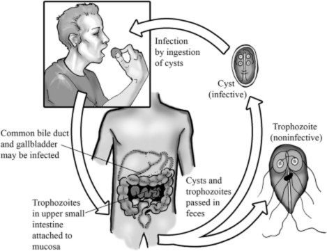
- Infection: The life cycle of an infection starts when the host is exposed to the infective version of the parasite. These could be trophozoites, cysts, or any other stage that is specialized. The method of transmission may differ, including the consumption of food items or water or direct contact with affected people, or transmission via vectors.
- Excystation: If the infective version of the parasite’s form is called a cyst it will undergo an process known as excystation before it can transform to the stage of trophozoite. The process occurs after the cyst has been subjected to conditions that are favorable like the right temperature, pH or the presence of certain chemicals or enzymes. This allows the cyst to split and release the trophozoites.
- Stage of the Trophozoite: After being exterminated, trophozoites transform into active and mobile versions from the parasite. They begin feeding, multiplying and could cause disease in the body of the victim. The trophozoite stage is usually closely associated with the invasive and pathogenic stage of the disease.
- Growth and reproduction: The growth of trophozoites within the host via a variety of methods, including binary fission and other methods of sexual reproduction. The process leads the host to a growth in the number of parasites and may cause destruction of the host’s tissues.
- Encystation: In a few parasites, the trophozoites undergo a process referred to as”encystation,” where they change into cysts. The process is usually caused by a variety of adverse circumstances like changes in the environment surrounding the host or the requirement to transfer to a different host. In the course of encystation the trophozoite goes through transformations in its morphology and forms the cyst’s protective surrounding it.
- Transmission: Cysts can be described as an type of parasite that is infectious that permits transfer to a different host. They can be eliminated by the infected host via excretions from the body, feces or via vectors. When they are in the outside environment the cysts may survive until they are consumed by the appropriate host, which can trigger an infection cycle.
- Environmental survival: The Cysts’ survival in the environment is crucial. They tend to be adapted to live in the outside environment which allows them to transfer their infection onto new hosts. They are able to endure harsh conditions like temperatures extremes, desiccation as well as exposure to disinfectants which improves their survival and the possibility of infectivity.
- Completed life cycle: The life cycle is complete when the ingested cysts, or the trophozoites are in the right host conditions and then transform into the stage that is required to replicate and cause pathogenesis.
It’s crucial to understand that the life-cycles of protozoan parasites may be complex, with various levels, hosts intermediate as well as vectors. The specifics of the life cycle including the length and character of each stage be different depending on the species of parasite. An understanding of the process as well as the development of protozoan parasites can be vital to ensure effective prevention, control and treatment strategies.
Diagnostic Techniques
Methods for diagnosing protozoan parasites such as the detection of cysts and trophozoites can differ depending on the particular parasite species and the location of infection.
Here are some commonly used methods used for the detection of parasitic infections:
- Microscopic examination: Microscopic examination of samples from clinical settings like feces blood, urine or tissue samples is an essential method of finding protozoan parasites. Slides of samples are made which are stained, then scrutinized under a microscope to see the distinctive characteristics of cysts and trophozoites. There are a variety of staining techniques like iodine and modified acid-fast or the trichrome staining method, can be used to increase the clarity of.
- Antigen detection:Antigen detection tests involve the identification of specific parasite antigens found in human samples such as blood or stool using techniques like enzyme-linked immunosorbent assay (ELISA), immunofluorescence assays or Rapid Diagnostic Tests (RDTs). Antigen detection provides fast and precise identification of certain protozoan parasites.
- A polymerase chain reaction (PCR): PCR (polymerase chain reaction) is a molecular method employed to detect and amplify genes (DNA or RNA) of protozoan parasites. Such tests can help differentiate and classify parasite types even at lower concentrations than conventional microscopic examination, making PCR tests particularly helpful where microscopic examination is difficult or not conclusive. Specific drug resistance markers or tracking response to treatment can also be determined with this test method.
- Serological tests: Serological methods use antibodies produced by the host as a response to parasite infection to detect current or past infections as well as determine their extent. They’re useful both in diagnosing current or past infections as well as measuring exposure levels of individual parasite species; most serological tests include ELISA assays, indirect immunofluorescence (IFA) tests as well as Western Blotting procedures.
- Image-based techniques: Imaging techniques such as ultrasound or computed tomography as well as magnetic resonance imaging, are sometimes utilized to assess and visualize tissue damage caused by protozoan parasites, particularly within organs like the spleen, liver or brain. Such tools have proven particularly successful at pinpointing infections linked to protozoans such as those residing therein.
- Culture and isolation: Isolating and cultivating parasites from clinical samples can help in their identification and characterization. This method involves incubating the specimen in specific conditions and growth media that encourage an increase in the size of the parasite. The process of culturing and isolating parasites can be essential for the production of viable trophozoites, or for tests to determine susceptibility to drugs.
- Molecular sequencing: Molecular sequencing methods that include the next generation sequencing (NGS), as well as Sanger sequencing, can be utilized to characterize and identify protozoan parasites on the basis of the genetic data they contain. These methods are particularly helpful to study genetic diversity and identifying emerging or new parasite species, and gaining an understanding of the molecular pathology that causes parasite diseases.
It is essential to speak with medical professionals or reference laboratory guidelines to determine the most appropriate test method depending on the parasite species that is suspected and the medical situation. The proper sample collection, transportation and processing techniques are vital to ensure precise and reliable results.
Summary and Conclusion
Cysts and trophozoites are two distinct phases within the protozoan lifecycle of parasites. Cysts are non-motile, dormant structures that have a protective outer membrane or wall and trophozoites, on the other hand, are actively mobility-based versions of the parasite, without any protective cover.
Cysts have no metabolic activity and are principally responsible for the transmission of disease and their survival beyond the host. They are adapted to allow them to endure harsh conditions in the environment and are therefore the pathogens that cause infection in the parasite. Cysts are frequently detected in diagnostic samples and indicate an infection.
Understanding the specific characteristics and behavior of trophozoites and cysts is vital for accurate diagnosis, proper treatments, as well as the design of measures to control specific phases of the parasite’s cycle. Further studies and research on specific species of parasites are required to gain a thorough knowledge of the many varieties of protozoan diseases and their medical significance.