Lagophthalmos and Ptosis are two distinct eyelid abnormalities that affect an individual’s ocular health and appearance. Lagophthalmos is characterized by the inability to close the eyelids completely, leaving parts of the eye exposed, which can potentially lead to complications like corneal damage. On the other hand, ptosis refers to the drooping or falling of the upper eyelid, often giving the individual a tired appearance and, in severe cases, impairing their field of vision. Both conditions can arise from a variety of causes, and understanding their differences is crucial for proper diagnosis and treatment.
What is Lagophthalmos?
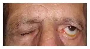
Lagophthalmos refers to the inability of an individual to close their eyelids completely, resulting in partial or total exposure of the eye. This condition can potentially lead to dryness, irritation, and other complications due to the exposed cornea and conjunctiva.
Causes of Lagophthalmos:
-
- Facial nerve paralysis (Bell’s palsy): The facial nerve controls various muscles around the face, including the orbicularis oculi muscle responsible for eyelid closure. Damage or paralysis of this nerve can cause the eyelid to not close fully.
- Scarring: Scarring from burns, trauma, or surgeries can affect the skin elasticity and prevent the eyelids from closing completely.
- Proptosis: This is the forward bulging of the eye, commonly seen in conditions like Graves’ disease. A bulging eye can prevent the eyelids from enveloping the eyeball fully.
- Previous surgeries or trauma: Surgeries or injuries to the eyelid or orbital area can sometimes lead to lagophthalmos.
- Congenital conditions: Some individuals are born with eyelids that cannot close completely.
Complications: The primary concern with lagophthalmos is the potential damage to the cornea due to its exposure. The cornea relies on regular blinking and a closed eyelid during sleep to maintain its moisture and health. In lagophthalmos, the constant exposure can lead to:
- Dryness
- Irritation
- Infections
- Ulceration of the cornea
- Visual disturbances
Treatment primarily focuses on protecting the exposed portion of the eye and might include:
- Lubricating eye drops and ointments
- Nighttime moisture chambers or goggles
- Surgical procedures, like tarsorrhaphy, to partially close the eyelids
- Eyelid weights to help the eyelids close
Early detection and appropriate treatment are crucial to prevent complications and maintain ocular health.
What is Ptosis?
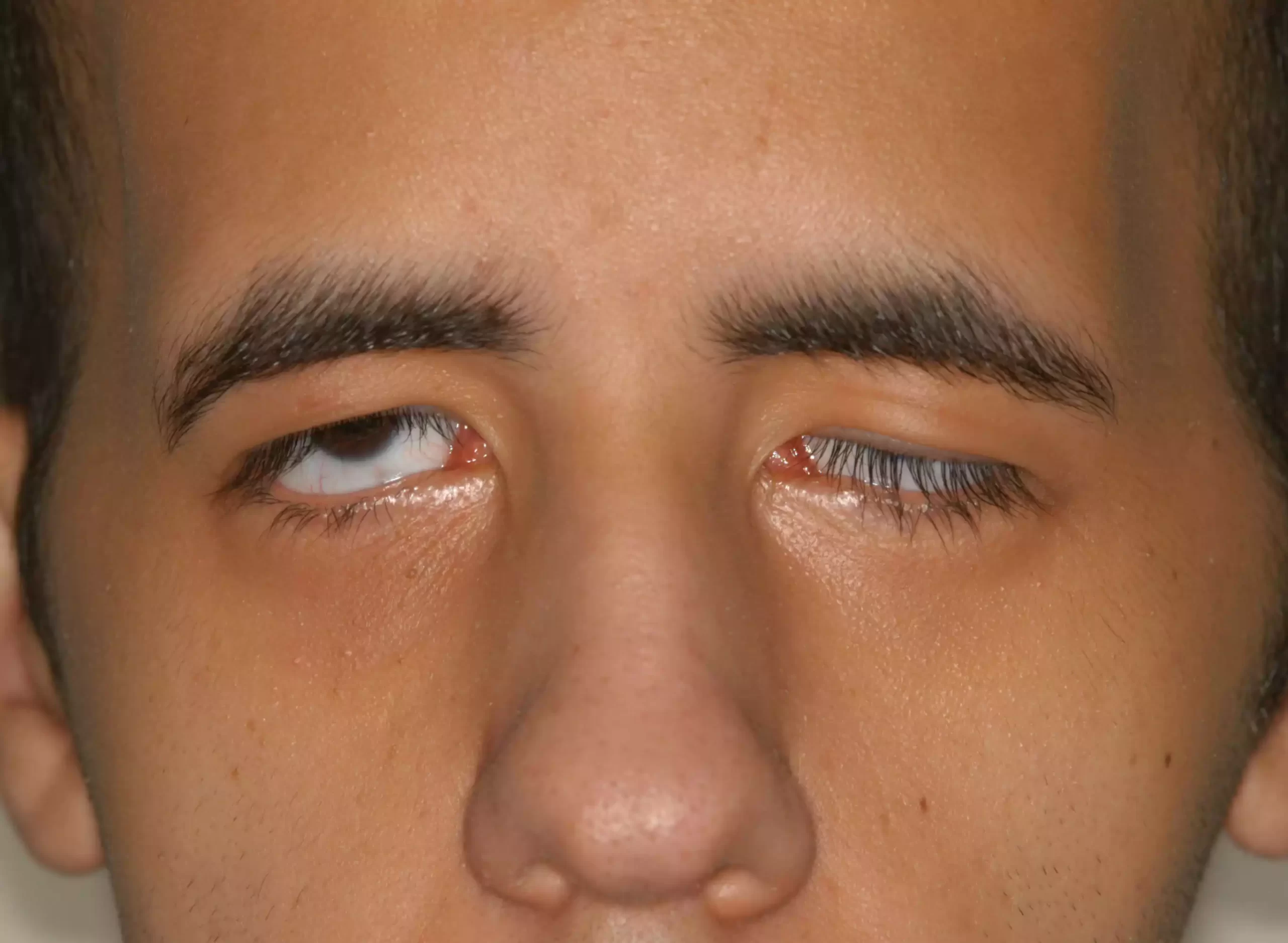
Ptosis (pronounced “toe-sis”) refers to the drooping or falling of the upper eyelid. This condition can affect one or both eyes and can range from being barely noticeable to covering the pupil entirely. Ptosis can impact not only an individual’s appearance but, in severe cases, also their vision.
Causes of Ptosis:
-
- Congenital: Some individuals are born with ptosis due to the underdevelopment of the levator muscle, which is responsible for lifting the upper eyelid.
- Age-related: As people age, the tendon that connects the levator muscle to the eyelid can stretch and weaken, leading to drooping.
- Neurological disorders: Certain conditions, such as Horner’s syndrome or third cranial nerve palsy, can cause ptosis.
- Myasthenia gravis: An autoimmune neuromuscular disorder that causes muscle weakness can affect the muscles responsible for elevating the eyelid.
- Trauma: Injuries to the eye or surrounding area can damage the muscles or tendons and lead to ptosis.
- Previous surgeries: Eye surgeries, especially those related to cataract or LASIK, can sometimes result in ptosis as a complication.
Complications: Ptosis can lead to:
- Amblyopia (lazy eye) in children, as the drooping eyelid can hinder proper visual development.
- Astigmatism or other refractive errors due to the altered eyelid position.
- Strabismus (misaligned eyes).
- Reduced visual field, especially if the drooping lid covers part of the pupil.
Treatment for ptosis depends on its cause and severity:
- Surgical correction is often the primary treatment, especially for congenital or age-related ptosis. The surgery aims to tighten or reposition the levator muscle or its tendon.
- Frontalis sling operation is used, particularly for cases where the levator muscle is profoundly weak. This procedure uses the forehead muscle (frontalis) to lift the eyelid.
- Medical treatments might be used for conditions like myasthenia gravis.
Regular check-ups and early intervention can help prevent complications and improve the cosmetic and functional outcomes for individuals with ptosis.
Comparison Table of Lagophthalmos and Ptosis
Below the comparison table between Lagophthalmos and Ptosis:
| Feature | Lagophthalmos | Ptosis |
|---|---|---|
| Definition | Inability to close the eyelids completely. | Drooping or falling of the upper eyelid. |
| Appearance | Eye remains partially or fully open. | Upper eyelid sags or droops downward. |
| Primary Affected Area | Eyelids | Upper eyelid muscle or its connecting tendon. |
| Common Causes | – Facial nerve paralysis (Bell’s palsy)
– Scarring – Proptosis (e.g., Graves’ disease) – Trauma or surgery |
– Congenital muscle weakness
– Age-related muscle or tendon weakening – Neurological disorders – Trauma |
| Potential Complications | – Dryness
– Corneal ulceration – Infections |
– Amblyopia (in children)
– Reduced visual field – Astigmatism |
| Treatment | – Lubricating eye drops and ointments
– Eyelid weights – Tarsorrhaphy (partial eyelid closure surgery) |
– Surgical correction of levator muscle
– Frontalis sling operation – Medical treatments for underlying causes |
This table provides a concise comparison of the primary features, causes, complications, and treatments for lagophthalmos and ptosis. It’s important to note that while this table highlights key differences and similarities, a comprehensive understanding would require a more in-depth exploration of each condition.
Diagnosis Techniques
The diagnosis techniques for both Lagophthalmos and Ptosis are crucial for proper treatment and management of the conditions. Here are the primary diagnostic methods used for each:
Lagophthalmos:
- Clinical Examination: The primary diagnostic method is the direct observation of incomplete eyelid closure.
- Snap Test: After asking the patient to blink, the time taken for the eyelid to return to its original position can provide insights into the laxity of the eyelid.
- Lagophthalmos Measurement: Using a ruler to measure the distance of the gap between the upper and lower eyelids.
- Rose Bengal or Fluorescein Staining: Helps in identifying any dry spots or abrasions on the cornea caused by exposure.
- Schirmer’s Test: Assesses tear production, which can be affected in cases of lagophthalmos.
- Electromyography (EMG): In cases where facial nerve dysfunction is suspected, this test evaluates the electrical activity of the involved muscles.
Ptosis:
- Clinical Examination: The degree of eyelid droop is evaluated by comparing the position of the eyelid to the pupil.
- Marginal Reflex Distance (MRD): Measures the distance between the center of the pupil and the eyelid margin.
- Levator Function Test: Assesses the function of the levator muscle by measuring the distance the eyelid can be lifted from its lowest to its highest position.
- Phenylephrine Test: A diagnostic test in which a drop of phenylephrine is placed in the eye to determine if there’s improvement in eyelid position. It helps differentiate between certain types of ptosis.
- Tension Tests: For suspected cases of myasthenia gravis, the ice test or Tensilon test might be used.
- Slit-lamp Examination: A detailed examination of the eyelid’s structure, which can help identify any anomalies or scars causing the ptosis.
- Neurological Examinations: If a neurological cause is suspected, tests like MRI, CT scans, or nerve conduction studies may be recommended.
Both conditions may require a combination of the above tests to arrive at a definitive diagnosis and to rule out underlying causes. It’s important for individuals with symptoms of either condition to consult with an ophthalmologist or oculoplastic surgeon for a thorough examination and appropriate diagnostic testing.
Recovery and Post-treatment Care
Recovery and post-treatment care are vital components of the treatment journey for both Lagophthalmos and Ptosis. Proper care ensures optimal healing, reduces the risk of complications, and can enhance the cosmetic and functional outcome of treatments.
- Lagophthalmos:
Recovery:
- Depending on the procedure, the recovery period can range from a few days to several weeks.
- For surgeries like tarsorrhaphy or gold weight implantation, complete healing might take a few weeks.
Post-treatment Care:
- Antibiotic Ointment: To prevent infections at the surgical site.
- Cold Compress: To reduce swelling and bruising in the initial days post-surgery.
- Avoid Rubbing or Touching the Eye: This minimizes the risk of infections and injury.
- Continue Using Lubricating Drops: To keep the eye moist and comfortable.
- Regular Follow-ups: To monitor healing and ensure that the treatment objectives are being met.
- Protection: Use sunglasses outdoors to protect the eyes from debris and excessive light.
- Ptosis:
Recovery:
- Post-surgical swelling and bruising are expected, usually peaking around 48 hours after surgery and then gradually diminishing over the following week.
- Most patients can return to non-strenuous activities within a week, but full recovery might take several weeks.
Post-treatment Care:
- Antibiotic Ointment: Typically applied to the surgical site to prevent infection.
- Cold Compress: Helps reduce swelling and discomfort for the first 24-48 hours.
- Eye Patch: Some patients might be required to wear an eye patch for a day or two to protect the surgical site.
- Avoid Strenuous Activities: Heavy lifting, bending, or vigorous exercises should be avoided for a few weeks post-surgery.
- Avoid Rubbing the Eye: This can interfere with the healing process.
- Continue Any Prescribed Medications: Especially if ptosis was due to an underlying medical condition.
- Regular Follow-ups: To monitor healing, check suture sites, and ensure the eyelid position is optimal.
- Protection: Wear sunglasses to protect the eyes from environmental irritants and sun exposure.
For both conditions, patients should be vigilant about any signs of complications, such as increased pain, excessive swelling, vision changes, or signs of infections like redness, warmth, or discharge. Prompt communication with the healthcare provider is vital for addressing any concerns during the recovery period.
Preventive Measures
Preventive measures for Lagophthalmos and Ptosis mainly revolve around avoiding factors that may lead to these conditions or managing underlying causes efficiently. Here are some preventive strategies for both:
Lagophthalmos:
- Eye Protection: Using protective eyewear during activities that pose a risk of trauma can prevent injuries that might lead to lagophthalmos.
- Manage Underlying Conditions: Regular monitoring and appropriate management of conditions like Graves’ disease can prevent the progression to lagophthalmos.
- Avoid Excessive Eye Surgeries: Multiple surgeries can increase the risk of complications, including lagophthalmos. Consultation with experienced surgeons and understanding potential risks are crucial.
- Facial Nerve Health: For those at risk of conditions like Bell’s palsy, taking precautions such as protecting against cold exposure can be beneficial.
- Healthy Lifestyle Choices: Regular eye check-ups and maintaining general health can help in early detection and management.
Ptosis:
- Avoid Trauma: Protecting the eyes from trauma by using protective eyewear during risky activities can prevent injury-related ptosis.
- Manage Medical Conditions: Conditions like diabetes or hypertension can impact eye health. Regular monitoring and appropriate management can reduce risks associated with these conditions.
- Regular Eye Check-ups: Early detection of any eye muscle issues can lead to timely interventions, potentially preventing the onset of ptosis.
- Understand Surgical Risks: Before undergoing any eye surgery, be aware of potential risks and complications. This awareness can help in making informed decisions and possibly choosing preventive strategies post-surgery.
- Promptly Address Neurological Symptoms: If experiencing symptoms like double vision, varying eyelid position, or rapidly changing vision, seek medical attention promptly. Early diagnosis and treatment can prevent conditions like myasthenia gravis from causing ptosis.
While some cases of lagophthalmos or ptosis (like congenital ones) may not be preventable, understanding potential risk factors and maintaining good ocular health can help in minimizing risks. Regular consultations with eye care professionals are vital for early detection and management.
Conclusion
Lagophthalmos and Ptosis are distinct ophthalmologic conditions impacting the eyelids, with the former resulting in the inability to close the eyelids completely, and the latter characterized by the drooping of the upper eyelid. While both conditions have different origins and presentations, their effective management is crucial to prevent further complications and to maintain visual function and ocular health. Diagnosis involves a range of clinical examinations and tests tailored to assess the respective features of each condition.
Treatment options are varied, including conservative management, surgical interventions, and treatments for underlying causes, with post-treatment care focusing on optimal recovery and prevention of complications. Finally, prevention is largely centered around protective measures, management of underlying health conditions, and maintaining eye health through regular check-ups and early interventions when abnormalities are detected.