Anencephaly and Microcephaly are congenital conditions affecting brain and skull development. Anencephaly is a severe neural tube defect where a baby is born without parts of the brain and skull, typically resulting in death shortly after birth. The exact cause is unknown, but it is associated with insufficient folic acid intake during pregnancy.
Microcephaly, on the other hand, is characterized by a significantly smaller head size due to abnormal brain development. Causes range from genetic mutations to maternal infections like the Zika virus. Its severity varies, with some children experiencing developmental delays or intellectual disabilities, while others have normal development.
What is Anencephaly?
Anencephaly is a serious developmental disorder categorized as a neural tube defect occurring when the upper part of the neural tube fails to close during embryological development. This condition results in the absence of major portions of the brain, skull, and scalp in newborns. Babies born with anencephaly are usually lacking significant parts of the brain responsible for thinking, hearing, vision, emotion, and coordination.
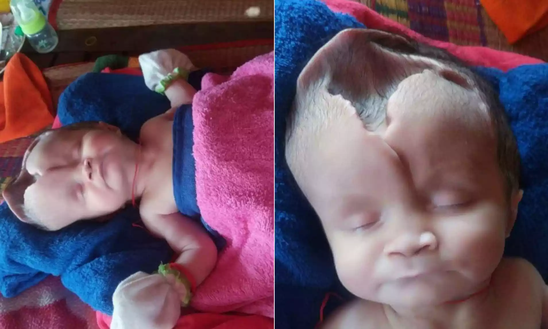
Unfortunately, this condition is usually fatal shortly after birth, with affected infants typically being stillborn or dying within hours to a few days after birth. The precise cause of anencephaly is unknown, but a deficiency in folic acid during pregnancy is considered a significant risk factor. Prenatal vitamins containing folic acid are recommended for women of childbearing age to reduce the risk of neural tube defects like anencephaly.
Causes of Anencephaly
Anencephaly is one of the more severe neural tube defects (NTDs). The exact cause of anencephaly, like most NTDs, is not completely understood and is likely multifactorial. Several potential causes and risk factors have been identified:
- Folic Acid Deficiency: A deficiency in folic acid (a type of B vitamin) before conception and during early pregnancy is a significant risk factor. Adequate intake of folic acid can help prevent neural tube defects.
- Genetic Factors: While anencephaly isn’t typically inherited in a simple pattern, families with a history of neural tube defects might have an increased risk.
- Environmental Factors: Exposure to certain environmental toxins may play a role.
- Medications: Some medications taken during pregnancy may increase the risk of neural tube defects.
- Maternal Diabetes: Uncontrolled diabetes in the mother can be a risk factor.
- High Body Temperature: Elevated body temperature (from fever or hot tub/spa use) in early pregnancy has been linked to an increased risk of anencephaly and other neural tube defects.
- Obesity: Maternal obesity, especially with a BMI of 30 or greater, increases the risk of having a baby with a neural tube defect.
- Race/Ethnicity: For reasons not fully understood, Hispanic mothers are at a higher risk for delivering babies with anencephaly compared to non-Hispanic white and black mothers.
Many cases occur without any identifiable risk factor. Preventive measures, particularly folic acid supplementation before conception and during early pregnancy, have been shown to significantly reduce the risk of anencephaly and other neural tube defects.
Symptoms of Anencephaly
Anencephaly is a severe congenital condition that results in underdevelopment of the brain and skull. The symptoms or manifestations of anencephaly are visually evident at birth. They include:
- Absence of the Skull: The most pronounced feature of anencephaly is the absence of a large part of the skull, particularly the bones that cover the front and top of the head.
- Absence or Underdevelopment of the Brain: Significant portions of the brain are missing, especially the cerebral hemispheres, which are responsible for thinking, vision, hearing, touch, and movement.
- Facial Feature Abnormalities: Despite the severe underdevelopment of the skull and brain, the facial features (such as the eyes, nose, and mouth) are usually present but might appear abnormally placed due to the missing portions of the skull.
- Heart Defects: Some babies with anencephaly might also have congenital heart defects.
- Lack of Reflexes: Due to the absence of significant portions of the brain, infants with anencephaly will not have typical reflexes such as sucking, reacting to touch, or responding to sounds.
Babies with anencephaly are usually blind, deaf, unconscious, and unable to feel pain. Regrettably, most infants with this condition are stillborn or die shortly after birth, although some may survive for a few hours or days. The condition can often be diagnosed before birth through prenatal testing such as ultrasound.
Treatment Options and Outlook
For anencephaly, the prognosis is extremely severe, and treatment options are limited. Here’s what you need to know:
Treatment Options:
- Palliative Care: There is no cure or standard treatment for anencephaly. Care is supportive and palliative, aiming to provide comfort to the newborn. This can include keeping the baby warm, administering medications to prevent discomfort or seizures, and providing gentle touch and care.
- Delivery Planning: Prenatal diagnosis can help healthcare providers and families prepare for the birth and make decisions about the management of the pregnancy and delivery. For instance, some families may choose to continue with the pregnancy and provide palliative care after birth, while others may consider the option of pregnancy termination.
- Organ Donation: In some jurisdictions and cases, families may consider the possibility of organ donation. This option can provide a means for the baby to give life to others, but the feasibility and ethical considerations vary by location and circumstance.
Outlook:
- Life Expectancy: Babies with anencephaly are often stillborn. Those who are born alive typically live only a few hours to a few days after birth. Prolonged survival is extremely rare.
- Emotional Impact: The diagnosis of anencephaly can be profoundly distressing for families. Grieving the loss of a child or managing the emotional impact of such a diagnosis is a deeply personal journey. Professional counseling and support groups can be invaluable in helping families navigate their grief and emotions.
Because of the severe nature of anencephaly and its associated prognosis, emphasis is often placed on prevention, primarily through folic acid supplementation before and during early pregnancy to reduce the risk of neural tube defects.
What is Microcephaly?
Microcephaly is a congenital condition characterized by a smaller than average head size when compared to babies of the same age and sex. This reduced head size is due to abnormal brain development either in utero or during the early years of life. In many cases, microcephaly is associated with intellectual and developmental disabilities, and it can also be accompanied by other neurological complications.

The severity and associated symptoms can vary widely among affected individuals. Causes of microcephaly can be genetic, environmental, or a combination of both, and can include factors such as infections during pregnancy, malnutrition, exposure to harmful substances, and certain genetic mutations.
Causes of Microcephaly
Microcephaly can result from a variety of causes, both genetic and environmental. Here are some of the key causes:
- Genetic Causes: Certain genetic mutations or disorders can lead to microcephaly. These can be inherited or occur spontaneously.
- Maternal Infections: Infections during pregnancy can affect fetal brain development. Notable infections include:
- Zika virus: A particularly well-publicized cause due to significant outbreaks in recent years and its clear link to microcephaly.
- Rubella (German measles)
- Cytomegalovirus
- Toxoplasmosis
- Others like syphilis, varicella-zoster (chickenpox), and certain types of herpes viruses.
- Severe Malnutrition: A lack of proper nutrients during pregnancy can hinder fetal brain growth.
- Exposure to Drugs and Toxins: Alcohol, certain drugs (both prescription and recreational), and toxic chemicals, when consumed or encountered during pregnancy, can interfere with fetal brain development.
- Interruption of Blood Supply: Conditions or complications during pregnancy that interrupt or reduce blood supply to the developing fetal brain can lead to microcephaly. This includes conditions like preeclampsia or certain types of trauma.
- Other Maternal Health Factors: Conditions like phenylketonuria (PKU) or uncontrolled diabetes in the mother can increase the risk of microcephaly.
- Interrupted Brain Development: Events that stop or slow the growth of the fetal brain can lead to microcephaly. This includes certain chromosomal abnormalities or congenital disorders.
- Craniosynostosis: This is a condition wherein the joints between the bony plates that make up the fetal skull close prematurely, preventing the brain from expanding and leading to microcephaly.
- Exposure to Radiation: High doses of radiation during pregnancy can affect fetal development.
The exact cause of microcephaly cannot be determined. Additionally, it’s possible for multiple factors to contribute simultaneously to the condition in certain individuals.
Symptoms of Microcephaly
Microcephaly is primarily characterized by a smaller than average head size. The severity of microcephaly and the range of associated symptoms can vary widely among individuals. Beyond the apparent reduced head circumference, the symptoms and complications can include:
- Developmental Delays: Children with microcephaly often experience delays in speech, movement, and other developmental milestones.
- Intellectual Disabilities: Many individuals with microcephaly have varying degrees of intellectual disability, ranging from mild to severe.
- Motor Function Issues: This can include challenges with balance and coordination.
- Facial Distortions: Due to the reduced size of the skull, some facial features may appear disproportionate.
- Seizures: A significant number of children with microcephaly experience seizures.
- Neurological Complications: This can include problems with vision, hearing, or swallowing.
- Hyperactivity: Some children with microcephaly may exhibit hyperactive behavior.
- Short stature: Overall reduced growth or dwarfism is sometimes associated with microcephaly.
- Difficulties with movement and balance: Motor problems may be present.
- Feeding problems: In severe cases, difficulties with feeding and swallowing can be evident.
All Individuals with microcephaly will experience these symptoms, and the severity can vary widely. Some children with milder forms of microcephaly might have a near-normal intelligence and not experience any significant developmental delays, while others with more severe forms may require intensive care and face lifelong challenges. As with many conditions, early intervention and appropriate supportive therapies can optimize outcomes and improve the quality of life for those affected.
Treatment and Management
Microcephaly is a lifelong condition with no cure. Treatment and management focus on addressing the symptoms and improving the quality of life for the affected individual. The specific interventions and therapies often depend on the severity of the condition and the particular challenges faced by the individual.
Here’s how microcephaly is typically managed:
- Early Intervention Services: For infants and toddlers, these services might include physical, occupational, and speech therapies to address developmental delays. Early intervention can be crucial in helping children reach their full potential.
- Medications: These can be used to treat symptoms and complications like seizures or hyperactivity.
- Physical Therapy: This helps improve motor function, strengthen muscles, and enhance balance and coordination.
- Occupational Therapy: This is beneficial for improving daily skills, such as feeding, dressing, and writing.
- Speech Therapy: It aids those with speech and language difficulties.
- Educational Support: Children with microcephaly may benefit from specialized educational programs tailored to their needs. This might include special education services or individualized education plans (IEPs).
- Routine Check-ups: Regular check-ups with a pediatrician can help monitor growth and development, and address any emerging health issues.
- Hearing and Vision Services: Regular screenings can detect issues related to hearing and vision, which are common in children with microcephaly. If problems are identified, early interventions like hearing aids or vision therapy can be beneficial.
- Supportive Care: Nutritional guidance, management of feeding difficulties, and other supportive measures can be essential, especially in severe cases.
- Counseling and Support: Families and caregivers might benefit from counseling to cope with the emotional and practical challenges of caring for someone with microcephaly. Support groups can also provide a valuable platform for parents to share experiences and advice.
- Genetic Counseling: If the cause of microcephaly is believed to be genetic, families might consider genetic counseling to understand the risks for future pregnancies.
Management requires a multidisciplinary approach involving a team of healthcare professionals, including pediatricians, neurologists, physical therapists, occupational therapists, speech therapists, and other specialists, depending on the needs of the individual.
Comparison Table of Anencephaly and Microcephaly
Here’s a comparison table outlining the differences between Anencephaly and Microcephaly:
| Aspect | Anencephaly | Microcephaly |
|---|---|---|
| Definition | A severe neural tube defect where major parts of the brain, skull, and scalp are absent. | A condition where the head (circumference) is significantly smaller than average due to abnormal brain development. |
| Causes | Often unknown, but associated with:
– Insufficient folic acid intake during pregnancy – Genetic factors – Environmental factors |
Can be due to a variety of factors including:
– Genetic mutations – Maternal infections (e.g., Zika virus) – Exposure to drugs or toxins during pregnancy – Severe maternal malnutrition – Interrupted brain development |
| Symptoms | – Absence of major parts of the brain and skull
– Facial features present but may appear abnormal – Often stillborn or live for a few hours/days |
– Small head size
– Developmental delays – Intellectual disabilities – Seizures, motor function issues, etc. |
| Treatment | No cure; focus is on palliative care | Focus on addressing symptoms and improving quality of life through therapies, medications, and supportive care. |
| Life Expectancy | Most infants die shortly after birth, typically within hours to days. | Varies widely; some individuals live into adulthood, depending on the severity and associated complications. |
While the table provides a concise overview, both conditions have complexities that go beyond this comparison, and individual cases can vary.
Prevention and Risk Factors
Both anencephaly and microcephaly have certain preventive measures and associated risk factors. Here’s a comparison:
Anencephaly
Prevention:
- Folic Acid Supplementation: One of the most effective prevention measures is taking folic acid before and during early pregnancy. Women of childbearing age are advised to take 400 micrograms (mcg) of folic acid daily.
Risk Factors:
- Folic Acid Deficiency: Insufficient intake before conception and during early pregnancy.
- Genetic Factors: Some families might have a higher risk.
- Environmental Factors: Exposure to specific toxins.
- Certain Medications: Use during pregnancy can increase the risk.
- Maternal Diabetes: Especially uncontrolled.
- High Body Temperature: Due to fever or external factors during early pregnancy.
- Obesity: Mothers with a BMI of 30 or greater.
- Race/Ethnicity: Higher incidence in Hispanic mothers compared to non-Hispanic white and black mothers.
Microcephaly
Prevention:
- Avoid Infections: Especially Zika virus, by using insect repellents, wearing protective clothing, and avoiding travel to affected areas during pregnancy.
- Avoid Alcohol and Drugs: Refrain from alcohol, illicit drugs, and certain medications not approved by a healthcare provider during pregnancy.
- Proper Nutrition: Ensure adequate nutrition, especially regarding folic acid intake.
- Control Chronic Conditions: Such as diabetes or phenylketonuria (PKU), through regular medical check-ups and following medical advice.
- Avoid Harmful Radiation: And toxins during pregnancy.
Risk Factors:
- Maternal Infections: Such as Zika virus, rubella, cytomegalovirus, and toxoplasmosis.
- Exposure to Drugs and Toxins: Alcohol, certain medications, and harmful chemicals during pregnancy.
- Severe Malnutrition: Inadequate nutrition during pregnancy.
- Genetic Factors: Certain genetic mutations or disorders.
- Other Maternal Health Factors: Conditions like uncontrolled diabetes or phenylketonuria (PKU).
It’s essential to understand that while preventive measures can significantly reduce the risk of these conditions, they can’t eliminate the risk entirely. Always consult with healthcare professionals about personal risks and appropriate preventive measures.
Importance of Prenatal Care
Prenatal care is vital for both the health of the mother and the developing fetus. It encompasses regular check-ups and interventions that help identify, prevent, and manage potential health issues. Here’s a breakdown of the importance of prenatal care:
- Early Detection of Complications: Through routine screenings and ultrasounds, potential issues like gestational diabetes, preeclampsia, and fetal growth restrictions can be detected early and managed.
- Fetal Health Monitoring: Regular ultrasounds can help ensure that the fetus is developing normally, check for congenital anomalies, and assess the position and health of the fetus.
- Nutrition Guidance: Healthcare providers can advise on proper nutrition to ensure that both mother and baby are getting essential nutrients. This includes guidance on folic acid intake, which reduces the risk of neural tube defects.
- Prevention of Birth Defects and Illnesses: Prenatal care includes screening for infections like rubella or toxoplasmosis, which can harm the fetus. Vaccinations, such as the flu shot and whooping cough vaccine, can protect both mother and baby.
- Medication Management: Providers can review any medications the pregnant individual is taking to ensure they are safe during pregnancy and make adjustments as necessary.
- Lifestyle Counseling: Healthcare providers can offer guidance on safe exercise, avoiding harmful substances (like alcohol, tobacco, and certain drugs), and managing stress during pregnancy.
- Emotional and Psychological Support: Pregnancy can bring about various emotional changes. Regular check-ins with healthcare providers offer opportunities to discuss feelings, concerns, and potential mood disorders like antepartum depression.
- Education: Mothers-to-be can learn about the stages of pregnancy, what to expect during labor and delivery, and basic newborn care.
- Preparation for Birth: Prenatal care can help mothers plan for labor and delivery, discussing pain relief options, birthing plans, and potential scenarios like c-sections.
- Postpartum Planning: Discussions about breastfeeding, contraception post-delivery, and what to expect in the weeks following birth can be addressed.
- Reduction of Neonatal and Maternal Mortality: Prenatal care plays a vital role in reducing the risk of death for both mothers and newborns by identifying and managing potential threats.
- Community and Resource Connection: Healthcare providers can connect expecting mothers with local resources, support groups, parenting classes, and more.
In essence, prenatal care is a holistic approach to ensuring the best possible outcomes for pregnancy, delivery, and the postpartum period. Regular attendance and active participation in prenatal care can greatly influence the health and well-being of both the mother and the baby.
Conclusion
Anencephaly and microcephaly are serious congenital neurological conditions marked by abnormal brain and skull development. Anencephaly is characterized by the absence of major parts of the brain, skull, and scalp, leading to almost certain neonatal death shortly after birth. Microcephaly, on the other hand, involves a significantly smaller head size due to underdeveloped brain structures, resulting in a range of potential developmental and neurological challenges that vary in severity.
The exact causes of these conditions can be multifaceted, encompassing genetic, environmental, and maternal health factors. While certain preventive measures, like folic acid supplementation, can reduce risk, there’s no cure for either condition. Prenatal care plays a crucial role in early detection and management.