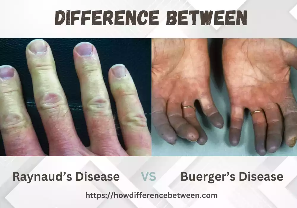
Raynaud and Buerger Disease both are distinct conditions in the medical field that impact the blood vessels and trigger similar symptoms. Both conditions are characterized by the constricting of blood vessels, they have distinct causes and impact different areas within the human body. In the article we’ll examine the main differences between Raynaud’s Disease and Buerger’s Disease, including their causes symptoms, signs, diagnosis and treatments.
Definition of Raynaud’s disease
Raynaud’s disease, commonly referred to as Raynaud’s Syndrome or simply Raynaud’s, is a medical disorder marked by periodic attacks of decreased blood flow to extremities – predominantly toes and fingers – usually the toes and fingers. This narrowing condition known as vasoconstriction typically manifests itself due to exposure to cold temperatures or emotional strain; its trigger can vary.
Raynaud’s Disease can be divided into two distinct forms; primary and secondary. Primary Raynaud’s, also referred to as Raynaud’s syndrome, does not have any apparent source and should be considered benign disease; in contrast secondary Raynauds often has links with other health conditions like Lupus (autoimmune lupus erythematosus arthritis rheumatoid) connective tissue diseases as well as certain medications or occupational hazards causing it.
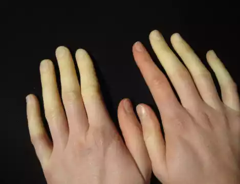
Raynaud’s Disease can occur anywhere on the fingers and toes due to reduced blood flow. When blood flow returns, areas affected may transition from blue to white in hue, before turning red when blood has returned; when restored again this could involve pain or tingling as the area can redden again with warmth; symptoms typically range from mild to serious depending on each individual and its frequency and duration can differ as well as frequency/duration variations over time.
Raynaud’s Disease affects women more frequently and typically manifests itself between early adolescence and adulthood, usually as early as during adolescence or even earlier. Although Raynaud’s can be annoying and disrupt daily activities, in most instances it doesn’t pose serious threats; proper management often relies on lifestyle modifications like keeping warm while reducing triggers while protecting extremities – in addition to medications designed to increase blood flow and alleviate attacks of symptoms.
Causes of Raynaud’s Disease
Raynaud’s Disease remains unexplained, its causes have yet to be fully identified; it is widely speculated as being due to a combination of environmental and genetic influences. An abnormal reaction by blood vessels when exposed to cold temperatures or emotional stress results in vasoconstriction that reduces blood flow causing vasoconstriction that limits its progress through vessels resulting in Raynaud’s Syndrome; other contributing factors may include decreased vitamin K intake in pregnant women or reduced levels of Vitamin D3.
For starters:
- Abnormal cardiovascular response: The blood vessels of the extremities, specifically toes and fingers, often respond excessively to certain triggers that stimulate them and result in excess contraction of these extremity arteries causing more contraction than necessary. The abnormal response could be the result of malfunction of both wall cellular structures within these blood vessels as well as muscles that regulate them contracting excessively.
- Dysregulation of the Nervous System: Our nerve system plays an integral part in controlling our response to stress and in controlling constriction of blood vessels; for patients suffering Raynaud’s Disease this system may become hypersensitive or oversensitive and cause excessive vasoconstriction.
- Genetic Factors: Evidence indicates that Raynaud’s Disease may contain genetic influences. It occurs frequently within families and suggests an underlying genetic predisposition; certain gene variants could impact severity and development of Raynaud’s.
- Secondary Raynaud’s: Raynaud’s disease may also arise as the result of existing medical issues or external triggers, like systemsic sclerosis (scleroderma), Lupus or Rheumatoid Arthritis – often with secondary causes including workplace hazards (ie using vibrating instruments), medications (beta-blockers & Ergotamine), chemical exposure or vinyl chloride exposure as possible culprits.
Understand the differences between primary Raynaud’s disease – where symptoms occur without an obvious source – and secondary Raynaud’s disease; both conditions share common triggers or sources. By knowing exactly why someone experiences Raynaud’s symptoms and the associated triggers and sources for them can manage and lessen them accordingly.
Symptoms of Raynaud’s Disease
Raynaud’s disease symptoms typically involve intermittent changes to appearance and temperature of affected areas – usually toes and fingers – caused by cold temperatures or emotional stress; they may vary in intensity or length over time, though.
Some prominent Raynaud’s Disease symptoms:
- Changes to Skin Color during: Episode The affected areas could become white (pallor) due to decreased blood flow. Once restored, those areas could change from pale (pallor) back into red (cyanosis) before finally becoming pinkish red (erythema) as blood vessels shrink back down again and begin constricting and relaxing as part of a pattern of constriction and relaxation of blood vessels and their consequences on color changes in affected areas.
- Cold Toes and Fingers: The affected areas typically appear extremely cold during an episode due to decreased blood flow or impaired circulation. This chilliness is due to reduced circulation.
- Tingling or Numbness: People suffering from Raynaud’s disease may experience the feeling of pins-and-needles sensation in affected toes, fingers or other parts of their limbs due to reduced oxygen supply to nerves due to reduced blood flow or temporarily stopped up blood vessels. The cause may include both temporary blood flow interruptions as well as reduced oxygen to nerve cells affecting them temporarily.
- Pain or discomfort: Sensations of pain or discomfort are never pleasant. After an incident occurs, sufferers may feel mild to moderate discomfort around their affected area ranging from throbbing pain to sharp piercing sensations that eventually subside as blood flow returns back to normal and circulation resumes its course.
- Skin Texture Alterations: In extreme cases, skin around affected areas could tighten and thin over time. Rarely ulcers or sores may form on toes and fingertips and this complication often appears with Raynaud’s diseases secondary to an autoimmune disease such as systemic sclerosis.
At times of Raynaud’s disease episodes, duration and intensity can differ for everyone involved. Some may experience short or occasional flare ups while other might experience more regular and lasting ones. Learning strategies that keep extremities warm and managing triggers could ease symptoms as well as improve quality of life for people living with Raynaud’s Disease.
Diagnosis of Raynaud’s Disease
As is generally accepted, diagnosis of Raynaud’s Disease generally entails both medical history analysis and physical exam/examined with diagnostic tests; typically performed by healthcare specialists like primary care doctors or rheumatologists; these steps typically are required in diagnosing Raynaud’s.
- Medical History: First, your doctor will conduct a complete medical history analysis that includes symptoms like frequency and duration as well as any trigger factors or patterns as well as existing medical conditions or medication that might play a part in Raynaud’s. This process helps distinguish secondary from primary Raynaud’s.
- Physical Examination: When conducting a physical exam, healthcare providers will closely inspect any affected fingers, toes or extremities for changes in color or any signs of ulcers or tissue damage, hotness in affected area as well as signs of any other abnormality.
- Test for Cold Stimulation: Sometimes called the cold test, this diagnostic tool could be administered in order to simulate Raynaud’s Coma. A doctor could submerge feet or hands into cold, icy water or expose them to an atmosphere with low temperatures while monitoring changes in color or blood vessel reactions; all while measuring whether this condition poses serious threats. This test can help diagnose your condition as well as ascertain its seriousness.
- Nailfold capillaroscopy: Nailfold capillaroscopy (NC) is a noninvasive imaging technique which involves studying small blood vessels at the base of fingernails through microscope. NC can help detect capillary abnormalities associated with autoimmune diseases that result in secondary Raynaud’s.
- Tests for blood: Tests designed to diagnose possible medical conditions that could contribute to secondary Raynaud’s may be necessary in order to assess any secondary Raynaud’s cases, including antinuclear antibody (ANA), erythrocyte sedimentation rate (ESR) or C-reactive proteins (CRP) levels in your system.
Diagnosing Raynaud’s disease typically relies on its typical symptoms and an extensive evaluation by medical specialists, along with diagnostic tests used to ascertain primary causes in secondary cases or situations of uncertainty about diagnosis. Early identification and management is important in order to avoid complications while improving quality of life for those living with Raynaud’s.
Treatment for Raynaud’s Disease
The treatment of Raynaud’s disease is to control and decrease the frequency and intensity of episodes, reduce complications, and increase circulation of blood to affected regions. The approach to treatment typically includes a mix of lifestyle changes and, in some instances medication. The treatment strategy may vary based on the intensity of the symptoms and if Raynaud’s is primary, or secondary.
Here are some typical treatments:
- Lifestyle modifications:
- Keep warm: Stay warm by dressing with layers, and wearing warm clothes particularly on cold winter day or during cold climates can prevent the development of. Be sure to protect the extremities by wearing warm socks, gloves and footwear that is insulated.
- Protect your feet and hands: Utilize foot and hand warming devices, warm gloves, socks and electric blankets for keeping affected regions warm.
- Avoid triggers: Find and stay clear of triggers that may trigger Raynaud’s symptoms like exposure to cold temperatures emotional stress, smoking.
- Stress management: Relaxation methods, such as mindfulness exercises that require deep breathing, mediation yoga, and meditation can lessen stress and decrease the chance of having attacks.
- Regular exercise: Regular physical activity can aid in improving circulation, and lessen the frequency of Raynaud’s attacks. But, it’s crucial to stay away from exercising in extreme cold weather.
- Medications:
- Calcium channel blockers: These medicines include nifedipine, amlodipine and diltiazem are typically prescribed to relax and expand blood vessels, thereby improving blood flow and reducing severity of Raynaud’s syndromes.
- Alpha-blockers: Medicines such as prazosin or doxazosin could be prescribed to ease the wall of blood vessels thus promoting greater blood flow.
- Topical treatments: The gel or cream can be applied to areas affected to aid in dilation of the blood vessels and increase blood flow.
- Other medicines: Sildenafil could be considered in cases where Raynaud’s disease is resistant or severe.
- Surgery:
- Surgery to treat sympathetic nerves: In a few instances when conservative treatment options aren’t effective, surgical procedures can be considered in order to cut off the sympathetic nerves controlling constriction of blood vessels. This procedure is generally reserved for the most severe cases and is performed by specialists.
It’s crucial for people suffering from Raynaud’s disease to collaborate with their physician to create a customized treatment program. Regular follow-up visits and continuous monitoring will help determine the efficacy of treatment and help make any needed adjustments.
Definition of Buerger’s Disease
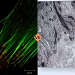
Buerger’s Disease, also referred to as thromboangiitis or obliterans, is an inflammatory disease with non-atherosclerotic features that typically impacts medium and small veins and arteries of extremities primarily feet and hands. Characterised by inflammation as well as blood clot formation inside blood vessels that create complete or partial obstruction leading to decreased flow to affected regions – which has strong connections with tobacco smoking as its root cause.
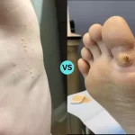
Buerger’s disease affects middle-aged to young adults between the ages of 20-40 years of age and more frequently among men. It manifests itself through symptoms including extreme pain in extremities with decreased or absent pulses; changed facial color (pallor, cyanosis or reddish tint), extreme cold sensitivity as well as ulcers or sores which do not heal; ulceration that does not close. If left untreated it could progress into gangrene which would require the affected limb(s) be removed altogether.

Buerger’s disease remains unexplained but is thought to involve an abnormal immune response triggered by tobacco toxins that stimulate inflammation and destruction of blood vessel walls, ultimately leading to inflammation, clotting in vessels, symptoms and complications characteristic of this condition.
Treating this condition requires quitting smoking completely as this habit worsens and increases risk for complications. Specific medications are administered to improve blood flow, reduce discomfort and stop formation of blood clots while in extreme cases bypass surgery may be needed to remove affected segments of vessels from circulation.
Regular follow up and management by an expert in treating Buerger’s Disease is necessary in order to halt its progress, relieve symptoms and limit complications.
Causes of Buerger’s Disease
Buerger’s disease (thromboangiitis Obliterans) remains unknown for certain. Several factors are believed to play a part. Smoking tobacco appears to be one of the primary contributors.
Other aspects associated with buerger’s disease development could include:
- Tobacco Consumption: Smoking cigarettes and using tobacco in general is a risk factor for Buerger’s disease, and avid or frequent smokers are especially at risk. Nicotine and other chemicals found in tobacco smoke directly damage blood vessel walls which then initiate an inflammatory response response from them and create inflammation within.
- Factors That Affect Immune Systems: Buerger’s Disease is characterized by an abnormal immune response. Researchers speculate that tobacco products act as triggers, initiating an inflammatory process in blood vessels which then causes formation of blood clots, swelling and an obstruction in circulation – leading to Buerger’s.
- Genetic Influences: Genetic factors could play a part in the formation of Buerger’s Disease. Studies suggest that certain genetic variants or immune system issues might increase an individual’s susceptibility. Its exact genetic causes remain unidentified.
- Environmental Factors: Smoking tobacco may play an integral role, but other environmental elements could contribute to its development as well. Exposure to certain chemicals found in industrial solvents or contaminated materials has been suggested as potential risk elements – although their role is yet to be fully evaluated in relation to Buerger’s Disease development.
Buerger’s disease typically impacts those aged 20-40 who smoke tobacco regularly – usually males. Ceasing to do so and managing or preventing its progression are both vital in managing this condition and managing Buerger’s progression. While its precise causes remain under study, smoking tobacco continues to play an instrumental part in its onset and progress.
Symptoms of Buerger’s Disease
Buerger’s disease, also referred to as thromboangiitis or obliterans, mostly affects blood vessels in the extremities. The disease is defined through inflammation, and development of blood clots in the medium and small veins and arteries, leading to decreased blood flow. The symptoms of Buerger’s Disease usually affect the limbs affected which are typically feet and hands.
These are the most prominent signs and symptoms that are associated with the Buerger’s Disease:
- The extremities are prone to pain: The most frequent characteristic of Buerger’s illness is pain, which usually is experienced in the hands as well as feet. The pain is typically described as a continuous, burning or throbbing sensation. It can get worse when you exercise or expose to cold temperatures, and can be relieved by rest.
- A decrease in blood flow: Buerger’s Disease causes obstruction or narrowing of blood vessels, which leads to diminished blood flow to affected regions. This may cause various signs and symptoms:
- Cold sensitivity: The feet and hands could be extremely cold or show an increased sensitization to cold temperatures.
- Changes in skin color: The skin of affected limbs could show changes in hue. It may appear white or pale (pallor) due to decreased blood flow. Then, it may change to blue (cyanosis) due to the effect of a lack of oxygenation. In certain cases it is possible for the skin to turn dark or red after blood flow is restored temporarily.
- Ulcers or sores: in the most severe cases, Buerger’s illness can result in the development of sores or ulcers that do not heal on the toes, fingers or on other extremities. The ulcers can be painful and can be caused by gangrene, tissue damage, or even gangrene.
- Puls that are absent or diminished: Buerger’s disease may cause the loss or absence of pulses in affected legs. The doctor may be unable to recognize pulses in a physical exam.
- Raynaud’s disease: A few people suffering from Buerger’s Disease may exhibit symptoms that are similar to Raynaud’s disease that include color changes as a result of extreme cold or stress. Raynaud’s syndrome in Buerger’s illness is typically more intense and persists for a long time.
Buerger’s illness typically manifests itself asymmetrically throughout the body; when one foot or hand has been affected, chances are its counterpart on the opposite side may also become susceptible. Over time, symptoms may worsen leading to severe complications like tissue damage, infections and even amputations; early diagnosis and prompt treatment are critical in order to stop its further progress and any associated consequences.
Diagnosis of Buerger’s Disease
Buerger’s disease (thromboangiitis Obliterans) requires a combination of clinical examination, medical history review and diagnostic tests in order to properly identify it. Diagnosis typically centers on its manifestation, such as when symptoms first appeared and type of involvement within blood vessels – as well as ruling out other illnesses with similar symptoms which might present with similar characteristics and timeframe.
Here are the techniques and procedures utilized during Buerger’s Disease diagnosis:
- Physical Examination and Medical History: A healthcare professional begins their assessment by gathering comprehensive patient health details as well as discussing smoking history and family histories of vascular illnesses. A thorough physical exam is then performed, specifically checking affected limbs for signs of reduced blood flow such as coolness or color changes, wounds or the presence of gangrene; additionally the doctor will check pulses in these affected areas.
- Tests for Blood: These blood-based exams are commonly undertaken to rule out other diseases and assess overall health in an individual, using techniques such as complete blood counts (CBC) or ESR tests and C-reactive proteins (CRP) analysis or Clotting Profile tests.
- Angiography: Angiography is a diagnostic imaging procedure which involves injecting contrast dye into blood vessels before taking X-ray pictures to identify them and any blockages, narrowing, or abnormal changes that might exist within them. As such, it has long been seen as the standard way of diagnosing Buerger’s disease.
- Doppler ultrasound: Doppler ultrasound utilizes sound waves to measure blood flow in affected legs. Doppler ultrasound helps assess arterial blockages as well as changes to vessel structures or response to stimuli like cold exposure by measuring their velocity over time. It allows physicians to quickly diagnose arterial issues.
- Imaging techniques: Other imaging tests such as magnetic resonance imaging (MRA) or computed tomography (CTA) may also be utilized to provide detailed images of blood vessels without resorting to any invasive procedures. These imaging studies offer more in-depth views into blood vessel health without necessitating additional procedures or invasive examination.
Buerger’s Disease can only be properly identified through ruling out other potential illnesses which produce similar signs, including atherosclerosis, autoimmune diseases and various vasculitis-related syndromes. A medical provider experienced in diagnosing and treating Buerger’s disease is essential in order to obtain an accurate diagnosis as well as appropriate therapy plans for its management.
Treatment for Buerger’s Disease
Buerger’s Disease, also referred to as thromboangiitis or obliterans and thromboangiitis obliterans, must be managed through treatment in order to halt its progress, relieve symptoms and avoid complications. A significant aspect of treatment should involve completely abstaining from tobacco as smoking cigarettes or drinking tobacco can aggravate symptoms and raise your chances of complications further.
Here is what makes up an effective plan of treatment:
- Smoking cessation: One key step toward managing Buerger’s disease is giving up cigarettes or any form of nicotine completely, whether by means such as counseling and replacement therapies or medication. Doing this can protect blood vessels further while improving overall outcomes. Healthcare professionals offer support to individuals through therapy sessions as they attempt to quit using nicotine products, including nicotine replacement therapies or prescription medication for this process.
- Medications:
- Vasodilators: Medicines that dilate blood vessels, including prostaglandin analogs, may help increase the flow of blood and relieve symptoms.
- Antiplatelet agents: Antiplatelet agents like Clopidogrel or Aspirin may be prescribed to decrease the chance of blood clot formation.
- Treatment Options for Pain: OTC or Prescribed pain relievers could help ease Buerger’s illness-induced discomfort.
- Wound Care: In cases of wounds and sores, appropriate wound care must be provided quickly and appropriately. This may involve regular cleansing with dressings to promote healing as well as specific products specifically made to address wound care needs. Surgical intervention might even be necessary in extreme circumstances to remove damaged tissues or treat non-healing wounds.
- Physical therapy and exercise: Regular exercises prescribed by medical professionals will improve circulation and relieve symptoms, while physical therapy exercises that increase range of motion or address any functional limitations caused by disease are suggested to increase range.
- Its Assistance measures:
- Maintain Warm Extremities: Protecting extremities that have been exposed to cold temperatures from vasoconstriction is critical in order to minimize symptoms and decrease vasoconstriction, so wearing warm clothing, heated socks or gloves, and staying away from cold environments is often advised as ways of keeping extremities warm.
- Care of Feet: Individuals living with Buerger’s diseases should take extra steps to maintain healthy feet in order to reduce complications, including receiving regular check-ups with proper hygiene measures and wearing footwear that improves circulation.
- Regular follow-up: Regular visits with a physician are crucial in monitoring disease progress and treatment effectiveness, and managing any issues or concerns that arise. Check-ups typically consist of evaluation of symptoms as well as physical exams or any necessary diagnostic testing procedures.
If the symptoms of Buerger’s Disease do not respond to conservative treatments, surgical options might be worth exploring. They might include bypass surgeries in order to reroute blood flow around blocked arteries or removal of affected segments from blood vessels.
Buerger’s illness sufferers must work closely with a medical specialist knowledgeable in managing this condition to achieve long-term control, prevent complications like gangrene or an amputation and ensure lasting control. Adherence to any treatment plans, lifestyle modifications and ongoing medical attention can ensure long-term control as well as ensure long-term survival of life without medical interventions such as an amputation.
Differences Between Raynaud and Buerger Disease
Raynaud’s Disease and Buerger’s Disease are two separate circulatory conditions that both impact extremities’ blood vessels. Both conditions can produce symptoms including pain and changes to color.
There are significant variations between them major ones being:
- Cause of the Problem:
- Raynaud’s Disease: Raynaud’s disease is a vasospastic condition characterised by episodes of excessive constriction of blood vessels in response to low temperatures or emotional strain. Although its exact causes remain enigmatic, some researchers suspect issues related to controlling the tone of your vascular walls could contribute.
- Buerger’s Disorder: Buerger’s Disorder is a non-atherosclerotic inflammatory disease characterized by formation of blood vessel clots and inflammation that obstruct blood circulation completely or partially; primary source is smoking cigarettes/tobacco products which leads to complete or partial obstruction in blood flow.
- Age and gender:
- Raynaud’s disease: Raynaud’s disorder typically strikes at younger age ranges – typically within their teens or early adulthood years – with female individuals more often affected than their male counterparts.
- Buerger’s Disease: Buerger’s illness typically impacts young and middle-aged adults between the ages of 20-40 years. Men tend to be affected more, and is also more prevalent among those who smoke tobacco products or take tobacco-containing products.
- Involvement in blood vessel systems:
- Raynaud’s disease: Raynaud’s illness affects small arteries that supply your toes and fingers, with occasional blood vessel spasms occurring which do not result in permanent tissue damage. These conditions tend not to result in long-term consequences but should only last up to 24 hours at most before healing occurs on its own.
- Buerger’s Disease: Its Buerger’s disease, commonly referred to as BUD, is characterized by inflammation and clot formation in small to medium sized veins and arteries running along extremities – specifically feet and hands – leading to blockages which block off blood vessels leading from them, potentially leading to injury to tissues, ulcers or in extreme instances leading to an infection known as Gangrene.
- Association with tobacco use:
- Raynaud’s Diseases: Raynaud’s illness does not directly relate to tobacco usage, and can occur even among people without history of tobacco usage or consumption.
- Buerger’s Disease: Berger’s illness has long been associated with smoking tobacco products; most individuals diagnosed with Berger’s illness are smokers or have extensive histories of smoking tobacco products. Treatment and Management Options for this Condition To reduce smoking-induced symptoms and manage Berger’s, treatment must address lifestyle habits which expose oneself to nicotine regularly – such as regular tobacco smokers who continue their habit or former heavy smokers with this illness may seek to stop, which are closely tied.
- Management and treatment:
- Raynaud’s Disease: Raynaud’s illness requires lifestyle modifications in order to eliminate triggers and increase circulation, including keeping extremities warm by limiting exposure to cold temperatures and managing stress effectively, keeping fingers warm with gloves on, as well as medication prescribed to relax blood vessels and increase flow.
- Buerger’s Disease: Treatment for Buerger’s disease focuses mainly on quitting tobacco usage altogether – such as cigarettes or chewing tobacco products. Smoking cessation or drinking tobacco consumption alone will significantly slow the progression of illness, while medication may help increase blood flow, ease discomfort and help avoid formation of blood clots; in extreme cases surgery could even be required to effectively address symptoms.
Although Raynaud’s and Buerger’s diseases differ substantially, there may be overlap in symptoms between both. Therefore, it may be possible for someone to suffer simultaneously from both conditions; an accurate medical assessment or diagnosis from a qualified expert is critical in order to ascertain which approach would work best to manage each illness effectively.
Final Thought
Raynaud’s Disease and Buerger’s Disease are two distinct illnesses that affect blood vessels, with a particular focus on the extremities. Raynaud’s disease manifests as instances of hyperintense blood vessel constriction due to stress or cold temperatures and Buerger’s disease is characterized by the development of clots and inflammation within the blood vessels. It is usually caused by smoking cigarettes.
Raynaud’s Disease typically begins around the age of teen or in the early years of adulthood. It most frequently affecting women. It mostly affects the arteries that are small in the toes and fingers. Buerger’s disease, on other hand, mostly affects older adults from middle age to young and, in particular, those who smoke, and affects the smaller and medium-sized blood vessels and veins in the feet and hands.
Although there are some differences between the two diseases but it’s crucial to recognize that symptoms can be overlapping, and some people may suffer from both Raynaud’s and Buerger’s disease. A medical professional must be sought out for a precise diagnosis and the appropriate treatment program.