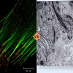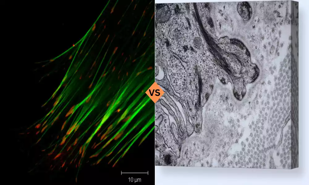
Focal Adhesion and Hemidesmosomes are two fundamental cell-matrix adhesion complexes that play crucial roles in connecting cells to their surrounding environment. Focal adhesions are dynamic structures found in various cell types, facilitating cell adhesion to the extracellular matrix. Comprising integrins, focal adhesion kinase (FAK), talin, and paxillin, they are central to processes such as cell migration, mechanotransduction, and signal transduction.
Hemidesmosomes are specialized adhesions primarily present in epithelial cells, particularly those lining body surfaces. They utilize α6β4 integrins, BP180, plectin, and laminin to anchor cells to the basement membrane, ensuring tissue integrity and barrier function. Understanding the distinctions and commonalities between these adhesions is vital for comprehending their significance in cell biology and human health.
What is Focal Adhesion?
Focal adhesions are dynamic multi-protein complexes that play a pivotal role in mediating cell adhesion to the extracellular matrix (ECM). They are typically found in adherent cells and are central to various cellular processes, including cell migration, mechanotransduction, and signal transduction. Focal adhesions are primarily composed of transmembrane proteins, intracellular signaling molecules, and cytoskeletal components. The key structural components of focal adhesions include integrins, focal adhesion kinase (FAK), paxillin, and talin.
Structure of Focal Adhesions
- Integrins: These are transmembrane proteins that span the plasma membrane of the cell, acting as the bridge between the ECM and the intracellular cytoskeleton. Integrins have an extracellular domain that binds to ECM proteins and an intracellular domain that interacts with cytoplasmic proteins.
- Focal Adhesion Kinase (FAK): FAK is a non-receptor tyrosine kinase that is localized to focal adhesions. It plays a crucial role in signal transduction by phosphorylating downstream targets in response to integrin activation.
- Talin: Talin is another cytoplasmic protein that links integrins to the actin cytoskeleton. It binds to the cytoplasmic tail of integrins, serving as a crucial mediator in the transmission of mechanical forces.
- Paxillin: Paxillin is an adaptor protein found in focal adhesions. It acts as a scaffolding protein, facilitating the assembly of signaling complexes and linking integrins to various signaling pathways.
Functions of Focal Adhesions
Focal adhesions are involved in several important cellular functions:
- Cell Adhesion: Their primary function is to anchor cells to the ECM, enabling stable attachment and migration.
- Mechanotransduction: Focal adhesions serve as mechanosensors, translating mechanical cues from the environment into intracellular signaling events.
- Signal Transduction: They play a crucial role in initiating intracellular signaling cascades, which can regulate processes such as cell growth, differentiation, and survival.
What is Hemidesmosomes?
Hemidesmosomes, like focal adhesions, are adhesive structures connecting cells to the ECM. They are structurally and functionally distinct. Hemidesmosomes are primarily found in epithelial cells, which line various surfaces in the body, including the skin and mucous membranes. These structures are critical for maintaining the integrity of epithelial tissues and are involved in adhesion to the basement membrane.
Structure of Hemidesmosomes
- Integrins: Similar to focal adhesions, hemidesmosomes also involve integrins, specifically α6β4 integrins. These integrins bind to laminin in the basement membrane.
- BP180 (Bullous Pemphigoid Antigen): BP180, also known as collagen XVII, is a transmembrane glycoprotein that connects integrins to the intermediate filament cytoskeleton. It plays a crucial role in stabilizing hemidesmosomes.
- Plectin: Plectin is a linker protein that connects the cytoplasmic domain of BP180 to intermediate filaments. This interaction provides structural stability to hemidesmosomes.
- Laminin: Laminin is a major component of the basement membrane and serves as the ligand for α6β4 integrins in hemidesmosomes.
Functions of Hemidesmosomes
Hemidesmosomes have several important functions, primarily in epithelial tissues:
- Epithelial Adhesion: They anchor the basal layer of epithelial cells to the basement membrane, which is essential for tissue integrity.
- Tissue Integrity: Hemidesmosomes play a vital role in maintaining the structural integrity of epithelial tissues, ensuring that they do not easily detach from the underlying matrix.
- Barrier Function: In tissues like the skin and mucous membranes, hemidesmosomes contribute to the barrier function, preventing the entry of pathogens and maintaining homeostasis.
Comparison Table of Focal Adhesion and Hemidesmosomes
To better understand the differences between focal adhesions and hemidesmosomes, let’s create a detailed comparison table:
| Characteristic | Focal Adhesions | Hemidesmosomes |
| Location | Found in adherent cells in various tissues. | Primarily found in epithelial cells, especially those lining body surfaces. |
| Composition | Comprised of integrins, focal adhesion kinase (FAK), paxillin, talin, and other signaling molecules. | Composed of α6β4 integrins, BP180 (collagen XVII), plectin, and laminin. |
| Primary Function | Anchoring cells to the extracellular matrix (ECM), facilitating cell migration, mechanotransduction, and signal transduction. | Anchoring the basal layer of epithelial cells to the basement membrane, maintaining tissue integrity, and contributing to the barrier function in epithelial tissues. |
| Mechanotransduction Role | Central in translating mechanical forces from the ECM into intracellular signaling events. | Also involved in mechanotransduction but with a focus on maintaining tissue integrity and barrier function. |
| Cell Type | Present in a wide range of cell types, including fibroblasts, myocytes, and endothelial cells. | Predominantly found in epithelial cells. |
| Structural Role | Act as connections between the ECM and the cytoskeleton, mainly the actin cytoskeleton. | Serve as connections between the ECM and intermediate filaments in the cytoskeleton. |
| Key Components | Integrins, FAK, talin, paxillin, actin, and various signaling molecules. | α6β4 integrins, BP180, plectin, laminin, and intermediate filaments. |
| Diseases Associated | Implicated in various diseases, including cancer metastasis, fibrosis, and cardiovascular diseases. | Often linked to skin diseases, such as epidermolysis bullosa and pemphigoid diseases. |
Similarities Between Focal Adhesion and Hemidesmosomes
While focal adhesions and hemidesmosomes have distinct structures and functions, there are some similarities between the two types of cell-matrix adhesions:
- Integrin Involvement: Both focal adhesions and hemidesmosomes rely on integrins for their connection to the extracellular matrix. In focal adhesions, integrins link cells to various ECM proteins, while in hemidesmosomes, they bind to laminin in the basement membrane.
- Cytoskeletal Linkage: Both structures involve the connection of integrins to the cytoskeleton. In focal adhesions, integrins link to the actin cytoskeleton through talin, while in hemidesmosomes, they connect to intermediate filaments via BP180 and plectin.
- Role in Mechanotransduction: Both focal adhesions and hemidesmosomes participate in mechanotransduction. They sense mechanical forces and transmit signals to the cell interior, which can result in various cellular responses.
- Cell Adhesion and Migration: Both structures are essential for cell adhesion and migration. Focal adhesions are critical for cell motility in various cell types, while hemidesmosomes play a role in the adhesion of epithelial cells to the basement membrane during tissue maintenance and repair.
Significance in Cell Biology
Understanding the differences and similarities between focal adhesions and hemidesmosomes is crucial for unraveling their significance in cell biology. These structures are involved in various physiological and pathological processes, and their dysregulation can lead to severe health conditions.
Focal Adhesions in Cell Biology
- Cell Migration: Focal adhesions play a pivotal role in cell migration, which is a fundamental process in embryogenesis, tissue repair, and immune responses. Dysregulation of focal adhesions can contribute to the invasion and metastasis of cancer cells.
- Signal Transduction: Focal adhesions are central hubs for signal transduction. They activate various signaling pathways, including those related to cell growth, differentiation, and survival. Aberrant focal adhesion signaling can lead to pathological conditions.
- Mechanotransduction: As mechanosensors, focal adhesions are critical for cells to respond to mechanical cues from the microenvironment. This process is essential in processes such as bone remodeling and cardiovascular physiology.
- Development: During embryonic development, focal adhesions are involved in processes like tissue morphogenesis, organogenesis, and neural crest cell migration.
- Disease Implications: Aberrations in focal adhesions are associated with various diseases, including cancer, fibrosis, cardiovascular diseases, and muscular dystrophies.
Hemidesmosomes in Cell Biology
- Epithelial Tissue Integrity: Hemidesmosomes are crucial for maintaining the structural integrity of epithelial tissues. Their role in anchoring the basal layer of epithelial cells to the basement membrane is vital for tissue homeostasis.
- Barrier Function: In epithelial tissues like the skin and mucous membranes, hemidesmosomes contribute to the barrier function, preventing the entry of pathogens and maintaining a protective barrier against environmental factors.
- Tissue Repair: Hemidesmosomes play a role in tissue repair and regeneration, especially in skin wounds and injuries.
- Disease Associations: Mutations or dysregulation of hemidesmosomal components are associated with skin diseases, including epidermolysis bullosa and pemphigoid diseases. These conditions result in blistering and skin fragility.
Conclusion
Focal Adhesions and Hemidesmosomes, though both serving as vital links between cells and their extracellular environment, exhibit distinct roles and structures. Focal adhesions are versatile and central to cell migration and signaling, while hemidesmosomes are specialized for anchoring epithelial tissues and maintaining their structural integrity. Understanding these differences is essential, as disruptions in these adhesions can lead to severe health conditions, highlighting their significance in cell biology and human health. Continued research in this field promises to unveil new insights into disease mechanisms and potential therapeutic interventions.